. 2014 Dec 16;111(50):E5383-92.
doi: 10.1073/pnas.1419553111. Epub 2014 Dec 1.

Recessive cardiac phenotypes in induced pluripotent stem cell models of Jervell and Lange-Nielsen syndrome: disease mechanisms and pharmacological rescue

Affiliations

Recessive cardiac phenotypes in induced pluripotent stem cell models of Jervell and Lange-Nielsen syndrome: disease mechanisms and pharmacological rescue

Miao Zhang et al. Proc Natl Acad Sci U S A. .

Abstract

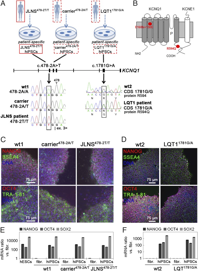
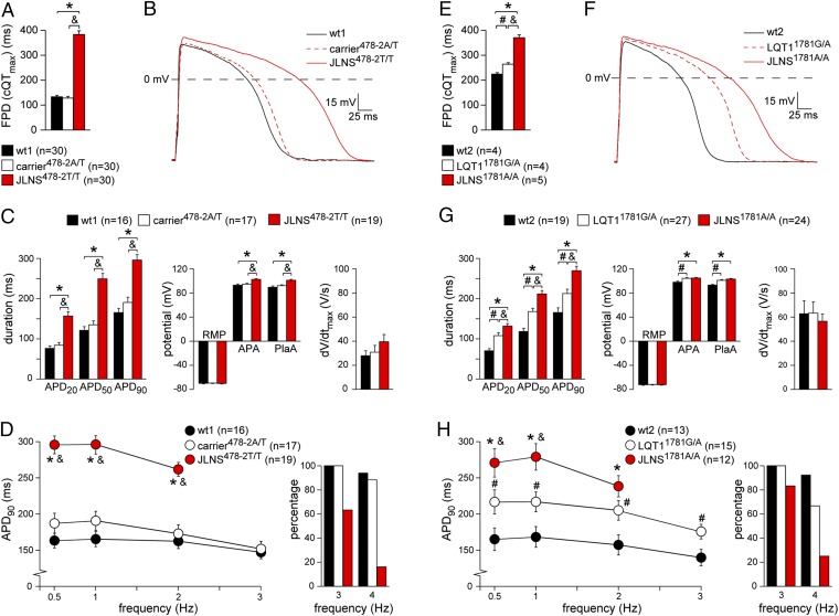
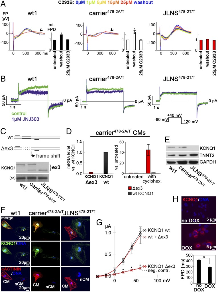
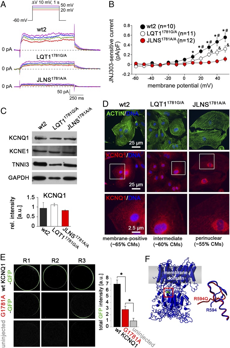
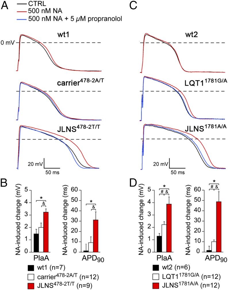
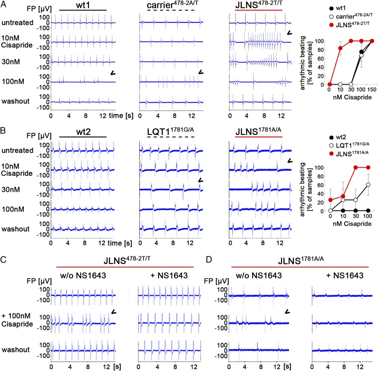
Jervell and Lange-Nielsen syndrome (JLNS) is one of the most severe life-threatening cardiac arrhythmias. Patients display delayed cardiac repolarization, associated high risk of sudden death due to ventricular tachycardia, and congenital bilateral deafness. In contrast to the autosomal dominant forms of long QT syndrome, JLNS is a recessive trait, resulting from homozygous (or compound heterozygous) mutations in KCNQ1 or KCNE1. These genes encode the α and β subunits, respectively, of the ion channel conducting the slow component of the delayed rectifier K(+) current, IKs. We used complementary approaches, reprogramming patient cells and genetic engineering, to generate human induced pluripotent stem cell (hiPSC) models of JLNS, covering splice site (c.478-2A>T) and missense (c.1781G>A) mutations, the two major classes of JLNS-causing defects in KCNQ1. Electrophysiological comparison of hiPSC-derived cardiomyocytes (CMs) from homozygous JLNS, heterozygous, and wild-type lines recapitulated the typical and severe features of JLNS, including pronounced action and field potential prolongation and severe reduction or absence of IKs. We show that this phenotype had distinct underlying molecular mechanisms in the two sets of cell lines: the previously unidentified c.478-2A>T mutation was amorphic and gave rise to a strictly recessive phenotype in JLNS-CMs, whereas the missense c.1781G>A lesion caused a gene dosage-dependent channel reduction at the cell membrane. Moreover, adrenergic stimulation caused action potential prolongation specifically in JLNS-CMs. Furthermore, sensitivity to proarrhythmic drugs was strongly enhanced in JLNS-CMs but could be pharmacologically corrected. Our data provide mechanistic insight into distinct classes of JLNS-causing mutations and demonstrate the potential of hiPSC-CMs in drug evaluation.

Keywords: Jervell and Lange-Nielsen syndrome; KCNQ1; disease modeling; human induced pluripotent stem cells; long QT syndrome.


Conflict of interest statement

Conflict of interest statement: C.L.M. is cofounder and advisor of Pluriomics.

Figures

Fig. 1.
Fig. 1.
Generation of hiPSCs from patients with KCNQ1 mutations. (A) Sequencing of the KCNQ1 gene identified the c.478-2A>T mutation at the splice acceptor site of intron 2 in the JLNS patient and the heterozygous carrier and the heterozygous c.1781G>A mutation in exon 15 of the LQT1 patient. The genotypes of the two healthy controls wt1 and wt2 are shown as reference. (B) The c.478-2A>T mutation is predicted to result in skipping of exon 3, with a concomitant reading frame shift from position 160 onwards, giving rise to a premature stop codon (E160fs+138X). The c.1781G>A mutation results in the substitution of an arginine with a glutamine at position 594 (R594Q) in the C-terminal domain of the KCNQ1 protein. (C and D) Immunofluorescence analysis of the pluripotency-associated markers NANOG, SSEA4, OCT4, and TRA1-81 in wt1, carrier478-2A/T, and JLNS478-2T/T (C), and in wt2 and LQT11781G/A hiPSCs (D). (E and F) qPCR expression analysis of the endogenous pluripotency genes NANOG, OCT4, and SOX2 in the indicated hiPSC lines.
Fig. 2.
Fig. 2.
Generation of isogenic JLNS hiPSC and hESC lines and general characterization of CMs. (A and B) CRISPR/Cas9-mediated homozygous disruption of the c.478-2 site in wt hESCs. Clones harboring frame shift-causing mutations on both alleles were isolated and expanded. (C and D) Schematic of the gene targeting strategy to introduce the c.1781G>A mutation in LQT11781G/A hiPSCs. Black boxes indicate exons (D). The wt allele of LQT11781G/A hiPSCs is shown with the black G in exon 15. The targeting vector has the mutation (A nucleotide, red) and a loxP-flanked G418-resistance cassette (NeoR). PCR primers a+b and c+d served to identify correctly targeted clones. (E and F) Immunofluorescence images of cardiac sarcomeric proteins MLC2a, MLC2v, TNNI, and α-actinin in the indicated lines following ∼4 wk of in vitro differentiation and maturation. F, Lower is a magnification of the framed area in Upper. (G and H) Heat map representation of qPCR gene expression of cardiac ion channels, before and after cardiomyocyte differentiation. Data are relative to wt hiPSCs. (I) Loss of KCNQ1 imprinting in wt1-CMs compared with undifferentiated hiPSCs. The SNP rs1057128 was used to discriminate between the two KCNQ1 alleles in wt1 cells. Data denote sequencing results of 20 RT-PCR clones.
Fig. 3.
Fig. 3.
Electrophysiological characterization of hiPSC-CMs. A–D show results for the wt1-, carrier478-2A/T-, and JLNS478-2T/T-CMs, and E–H for the wt2-, LQT11781G/A-, and JLNS1781A/A-CMs. (A and E) FPD quantification on MEAs. (B and F) Representative APs at 1 Hz. (C and G) Average APD20, APD50, APD90, dV/dtmax, RMP, APA, and PlaA. (D and H) Frequency dependence of APD90 (Left). Most JLNS-CMs could not be paced at high frequencies (Right). Symbols *, &, and # indicate statistical significance based on pairwise comparisons (P < 0.05).
Fig. 4.
Fig. 4.
Molecular mechanism underlying the c.478-2A/T mutation. (A) Effect of the IKs-blocker C293B on FPD. Single representative FPDs and average quantification are shown (n = 3). JLNS478-2T/T-CMs are insensitive to the drug. (B) Representative current traces before (control) and after JNJ303 treatment in the indicated hiPSC-CMs. IKs is virtually absent in JLNS478-2T/T-CMs. (Inset) Voltage protocol. (C) KCNQ1 RT-PCR analysis in the indicated hiPSC-CMs. (Top) Schematic showing primers used to reveal the skipping of exon 3 in mutant hiPSC-CMs. (Bottom) Upper band, wt transcript; lower band, Δex3 mRNA; pc, positive control. (D) qPCR analysis of relative abundance of Δex3 and wt KCNQ1 transcripts in carrier478-2A/T-CMs under baseline conditions (Left) and upon cycloheximide treatment (Right); cycloheximide (30 µg/mL, 3 h) selectively induces the Δex3 transcript. (E) Western blot analysis in hiPSC-CMs by using an antibody detecting the C terminus of KCNQ1. TNNT2 and GAPDH are shown as cardiac and loading controls, respectively. (F) Immunofluorescence analysis of KCNQ1 and α-actinin in hiPSC-CMs. Noncardiomyocyte cells are shown next to CMs to indicate specificity of staining. (G) Average I-V relationship for IKs measured upon injection of wt, Δex3, and wt+Δex3 mRNA into Xenopus oocytes (n = 10 each). (H) Genetic rescue of JLNS478-2T/T-CMs by using DOX-inducible KCNQ1 overexpression (Top); FPD shortening after 48 h of DOX treatment (Bottom, n = 4).
Fig. 5.
Fig. 5.
Molecular mechanism underlying the c.1781G>A mutation. (A) Representative IKs measured as JNJ303-sensitive current in the indicated hiPSC-CMs. (Inset) Voltage protocol. (B) Average JNJ303-sensitive tail currents in hiPSC-CMs. Symbols *, &, and # indicate statistical significance based on pairwise comparisons (P < 0.05). (C) Western blot analysis of KCNQ1 and KCNE1 in hiPSC-CMs (Top). TNNI3 and GAPDH are used as cardiac and loading controls, respectively. (Bottom) Densitometric quantification of the KCNQ1 band, normalized to TNNI3 (n = 2). (D) Immunofluorescence analysis of KCNQ1 and actin in hiPSC-CMs. Bottom are a magnification of the area framed in Middle. Percentages of CMs showing the indicated staining patterns are indicated. (E) Representative confocal images showing membrane localization of KCNQ1 following injection of GFP-tagged wt and G1781-mutated KCNQ1 mRNA into Xenopus oocytes (Left). Image-based quantification of total GFP intensity at the outer cell membrane (Right, n = 10 each). (F) Structural model of KCNQ1 highlighting the position of the R594 residue in the C-terminal assembly domain (Left) and change in predicted local structure induced by the R594Q mutation (Right).
Fig. 6.
Fig. 6.
Adrenergic stress and β-block in hiPSC-CMs. A and B show results for the wt1-, carrier478-2A/T-, and JLNS478-2T/T-CMs; and C and D for the wt2-, LQT11781G/A-, and JLNS1781A/A-CMs. (A and C) Representative APs at 1 Hz in presence of noradrenaline (NA, red) or NA+propranolol (blue). (B and D) Average PlaA and APD90 changes in presence of NA. Symbols *, &, and # indicate statistical significance based on pairwise comparisons (P < 0.05).
Fig. 7.
Fig. 7.
Drug-induced arrhythmia phenotypes in JLNS-CMs and rescue by a hERG activator. (A and B) Cisapride causes arrhythmias in spontaneously beating wt1-, carrier478-2A/T-, and JLNS478-2T/T-CMs (A) as well as in LQT11781G/A- and JLNS1781A/A- CMs. (B, Left) Representative MEA traces. Arrowheads indicate first appearance of arrhythmia. (B, Right) Statistics of arrhythmic beatings at different Cisapride doses from independent experiments (n = 2–6). (C and D) Pretreatment with the hERG channel activator NS1643 (30 µM) protects JLNS478-2T/T- (C) and JLNS1781A/A-CMs (D) from high-dose Cisapride-induced arrhythmia. Representative MEA traces from independent experiments are shown.

Comment in

References

    1. Jervell A, Lange-Nielsen F. Congenital deaf-mutism, functional heart disease with prolongation of the Q-T interval and sudden death. Am Heart J. 1957;54(1):59–68. - PubMed
    1. Schwartz PJ, et al. The Jervell and Lange-Nielsen syndrome: Natural history, molecular basis, and clinical outcome. Circulation. 2006;113(6):783–790. - PubMed
    1. Jespersen T, Grunnet M, Olesen SP. The KCNQ1 potassium channel: From gene to physiological function. Physiology (Bethesda) 2005;20:408–416. - PubMed
    1. Schulze-Bahr E, et al. KCNE1 mutations cause jervell and Lange-Nielsen syndrome. Nat Genet. 1997;17(3):267–268. - PubMed
    1. Romano C, Gemme G, Pongiglione R. [Rare cardiac arrythmias of the pediatric age. Ii. syncopal attacks due to paroxysmal ventricular fibrillation. (Presentation of 1st case in Italian pediatric literature)] Clin Pediatr (Bologna) 1963;45:656–683. - PubMed

Publication types

MeSH terms

Substances