. 2014 Dec 1;24(23):2861-7.
doi: 10.1016/j.cub.2014.10.029. Epub 2014 Nov 20.

The yeast polo kinase Cdc5 regulates the shape of the mitotic nucleus

Affiliations

The yeast polo kinase Cdc5 regulates the shape of the mitotic nucleus

Alison D Walters et al. Curr Biol. .

Abstract

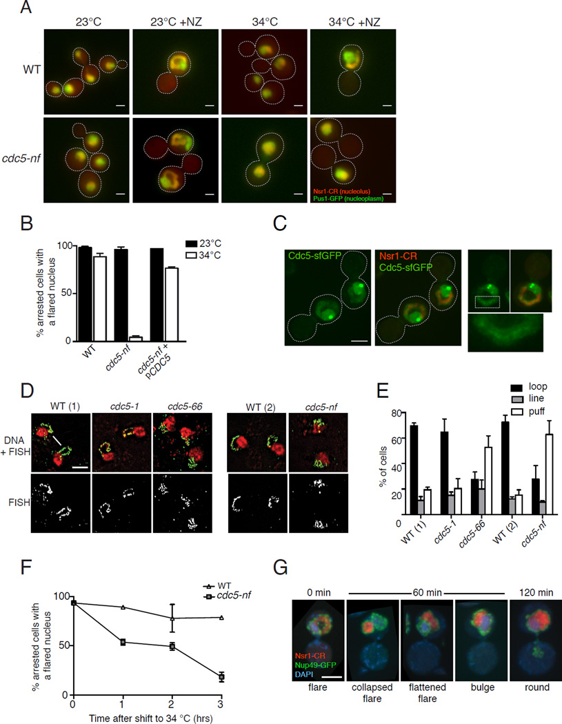
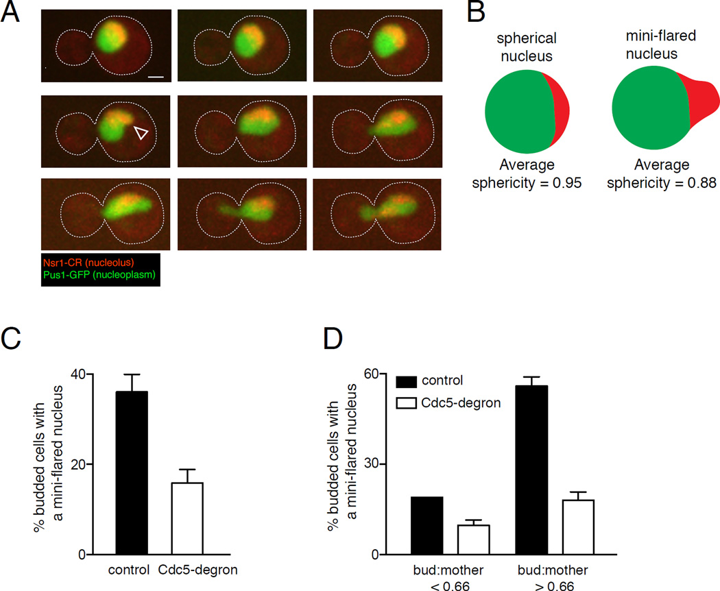
Abnormal nuclear size and shape are hallmarks of aging and cancer. However, the mechanisms regulating nuclear morphology and nuclear envelope (NE) expansion are poorly understood. In metazoans, the NE disassembles prior to chromosome segregation and reassembles at the end of mitosis. In budding yeast, the NE remains intact. The nucleus elongates as chromosomes segregate and then divides at the end of mitosis to form two daughter nuclei without NE disassembly. The budding yeast nucleus also undergoes remodeling during a mitotic arrest; the NE continues to expand despite the pause in chromosome segregation, forming a nuclear extension, or "flare," that encompasses the nucleolus. The distinct nucleolar localization of the mitotic flare indicates that the NE is compartmentalized and that there is a mechanism by which NE expansion is confined to the region adjacent to the nucleolus. Here we show that mitotic flare formation is dependent on the yeast polo kinase Cdc5. This function of Cdc5 is independent of its known mitotic roles, including rDNA condensation. High-resolution imaging revealed that following Cdc5 inactivation, nuclei expand isometrically rather than forming a flare, indicating that Cdc5 is needed for NE compartmentalization. Even in an uninterrupted cell cycle, a small NE expansion occurs adjacent to the nucleolus prior to anaphase in a Cdc5-dependent manner. Our data provide the first evidence that polo kinase, a key regulator of mitosis, plays a role in regulating nuclear morphology and NE expansion.


Figures

Figure 1
Figure 1. Cdc5 affects nuclear morphology during a mitotic arrest
A) Merged fluorescence images of fixed mitotic-arrested WT (top row) and cdc5-nf (bottom row) cells. The nucleoplasm is marked with Pus1-GFP (green) and nucleolus with Nsr1-mCherry (red). A nuclear flare containing the nucleolus forms in WT cells during a mitotic arrest induced by nocodazole (NZ) at both 23°C and 34°C (panels 2, 4) and in cdc5-nf cells at 23°C (panel 6). Nuclei of cdc5-nf cells are round at 34°C in the presence of NZ (panel 8). In the absence of NZ, cdc5-nf cells arrest in late telophase at 34°C (panel 7, Fig. S2E). White arrows indicate the interface between the nucleolus and nucleoplasm (panels 1 and 2). Scale bar, 2 µm. B) Quantification of phenotypes of NZ-treated cells shown in panel A (for each condition n=100 in at least 2 biological replicates). C) Cdc5 nuclear localization during a mitotic arrest. WT cells expressing Cdc5-sfGFP and Nsr1-mCherry (nucleolar marker) were arrested in NZ at 30°C and imaged using fluorescence confocal microscopy. Bright green spots are at the spindle pole bodies. Area contained in the white dashed box (right panels) is expanded in the bottom right panel to show a thin line of Cdc5-sfGFP within the nucleolar flare. Scale bar, 3 µm. D) Images from fluorescence in situ hybridization (FISH) using a probe for rDNA in WT and cdc5 mutants. Cells were arrested in NZ at 34°C for 150 min. WT(1) is isogenic to strains cdc5-1 and cdc5-66 while WT(2) is isogenic to strain cdc5-nf. WT cells arrested in mitosis typically exhibit an rDNA “loop” (arrow), while mutants defective in rDNA condensation exhibit amorphous structures referred to as “puff” (arrowhead) [31]. Scale bar, 5 µm. E) Quantification of rDNA phenotypes of cdc5 mutants from FISH described in D. F) Cdc5 is required for the maintenance of a nuclear flare. cdc5-nf and WT cells were arrested in mitosis at 23°C to allow flare formation and then shifted to 34°C. Samples were taken at the indicated time points. For each time point n=100 in at least 3 biological replicates. See also Fig. S2F. G) Images of cdc5-nf cells from the experiment in Fig. S2F showing examples of the collapse and eventual loss of nuclear flares upon the inactivation of Cdc5. NZ-arrested WT and cdc5-nf cells were shifted from 23°C to 34°C. Cells were fixed every 30 min and were imaged by confocal fluorescence microscopy. The images shown are from 60 and 120 min. The NE is marked with Nup49-GFP (green), the nucleolus with Nsr1-mCherry (red) and DNA with DAPI (blue). Scale bar, 3 µm. Error bars in all panels indicate SD.
Figure 2
Figure 2. Cdc5 is not affecting nuclear morphology through rDNA condensation or attachment to the NE
A) Images from FISH using a probe for rDNA in condensin mutants. Cells were arrested in NZ at 34°C for 150 min. rDNA is shown in green, DNA in red. Scale bar, 5 µm. B) Quantification of rDNA phenotypes of condensin mutants from FISH described in A. C) Merged fluorescence images of WT and condensin mutant cells arrested in NZ at 34°C. The nucleoplasm is marked with Pus1-GFP (green) and nucleolus with Nsr1-mCherry (red). D) Quantification of nuclear phenotypes for NZ-arrested condensin mutants experiment shown in C. WT(1) is isogenic to strain cdc5-nf while WT(2) is isogenic to strains brn1-9 and ycs4-1. E) Merged fluorescence images of asynchronous and NZ-arrested cells in a strain where the chromosomal rDNA was replaced by a single plasmid-borne copy of the rDNA. White arrows indicate nuclear flares. The nuclear pore subunit Nup49-GFP marks the NE (green), Nsr1-mCherry the "dot" nucleolus (red spot) and DAPI-stained DNA is shown in blue. F) Quantification of nuclear phenotypes of NZ-arrested rDNA-NE detachment mutants alone and in combination with the cdc5-nf allele. For panels B, D and F for each condition n=100 in each of at least 2 biological replicates. Error bars in all panels indicate SD.
Figure 3
Figure 3. Nuclei of cdc5-nf cells expand isometrically during a mitotic arrest
A) Outline of the experiment. WT and cdc5-nf cells were arrested in G1 with alpha factor and then released into NZ at 34°C. Samples were taken every 20 min and processed for soft X-ray imaging. B) Surface rendered views of the cell (pink) and nucleus (pale green) after segmentation of three dimensional reconstructions of WT and cdc5-nf cells from the experiment described in panel A. Scale bar, 1 µm. C) Quantification of nuclear surface areas as a function of cell size of WT and cdc5-nf cells (n=74 and 86, respectively). Nuclear flares begin to appear in cells of approximately 140 μm3 (black triangle). Linear regressions were calculated using Prism (R2 WT=0.633, R2 cdc5-nf=0.774). There is no significant difference between the slope of the line describing the increase in nuclear surface areas in WT and cdc5-nf (p=0.44, ANCOVA). Nuclei of WT cells have a slightly greater surface area than nuclei of cdc5-nf across all cell sizes (p=0.0006, ANCOVA). D) Quantification of nuclear volumes as a function of cell size of the same cells as panel C. There is no significant difference between nuclear volumes in WT (R2=0.677) and cdc5-nf (R2=0.762) across all cell sizes (p=0.48, ANCOVA).
Figure 4
Figure 4. Cdc5 allows NE expansion at the nucleolar region during an uninterrupted mitosis
A) Time course of images of a WT cell progressing from a small budded stage into anaphase. The nucleoplasm is marked with Pus1-GFP (green) and the nucleolus with Nsr1-mCherry (red). Images shown are maximum projections of merged fluorescence confocal images. A mini-flare (white arrowhead) is visible at 15 min. Scale bar, 1 µm. B) A mini-flare disrupts the spherical surface of the nucleus (green), specifically at the site of the nucleolus (red). Sphericity data are from Fig. S4A. C) Mini-flares are Cdc5-dependent. Quantification of the frequency of mini-flares in a control strain and Cdc5-degron cells. Fixed log-phase cells were imaged by fluorescence confocal microscopy and nuclear phenotypes of all budded cells were scored. The frequency of mini-flares in budded cells was lower in the Cdc5-degron strain (p<0.0001, Fisher’s exact test). D) Data from panel C divided into categories by bud:mother cell size ratio. For bud:mother <0.66, n=79 and 66 for WT and Cdc5-degron, respectively. For bud:mother >0.66, n= 69 and 134 for WT and Cdc5-degron, respectively. The frequency of mini-flares in cells with larger buds is significantly lower in the Cdc5-degron strain (p<0.0001, Fisher’s exact test). The difference in frequency of mini-flares in cells with small buds is not statistically significant (p=0.1665, Fisher’s exact test). Error bars in all panels represent SD.

References

    1. Chow K-H, Factor RE, Ullman KS. The nuclear envelope environment and its cancer connections. Nat. Rev. Cancer. 2012;12:196–209. - PMC - PubMed
    1. Scaffidi P, Misteli T. Lamin A-dependent nuclear defects in human aging. Science. 2006;312:1059–1063. - PMC - PubMed
    1. Kutay U, Hetzer MW. Reorganization of the nuclear envelope during open mitosis. Current Opinion in Cell Biology. 2008;20:669–677. - PMC - PubMed
    1. Witkin KL, Chong Y, Shao S, Webster MT, Lahiri S, Walters AD, Lee B, Koh JLY, Prinz WA, Andrews BJ, et al. The budding yeast nuclear envelope adjacent to the nucleolus serves as a membrane sink during mitotic delay. Curr. Biol. 2012;22:1128–1133. - PMC - PubMed
    1. Archambault V, Glover DM. Polo-like kinases: conservation and divergence in their functions and regulation. Nat Rev Mol Cell Biol. 2009;10:265–275. - PubMed

Publication types

MeSH terms

Substances