. 2014 Nov 17;24(22):2643-51.
doi: 10.1016/j.cub.2014.09.072. Epub 2014 Oct 16.

A comprehensive biophysical description of pairwise epistasis throughout an entire protein domain

Affiliations

A comprehensive biophysical description of pairwise epistasis throughout an entire protein domain

C Anders Olson et al. Curr Biol. .

Abstract

Background: Nonadditivity in fitness effects from two or more mutations, termed epistasis, can result in compensation of deleterious mutations or negation of beneficial mutations. Recent evidence shows the importance of epistasis in individual evolutionary pathways. However, an unresolved question in molecular evolution is how often and how significantly fitness effects change in alternative genetic backgrounds.

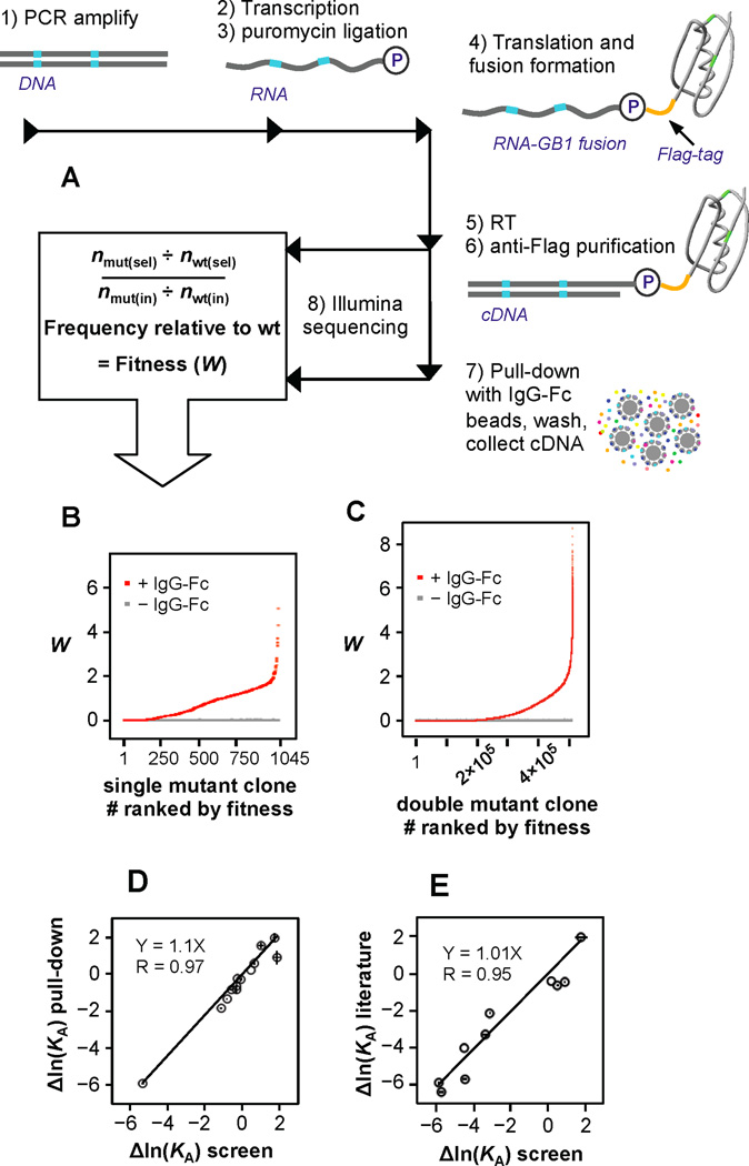
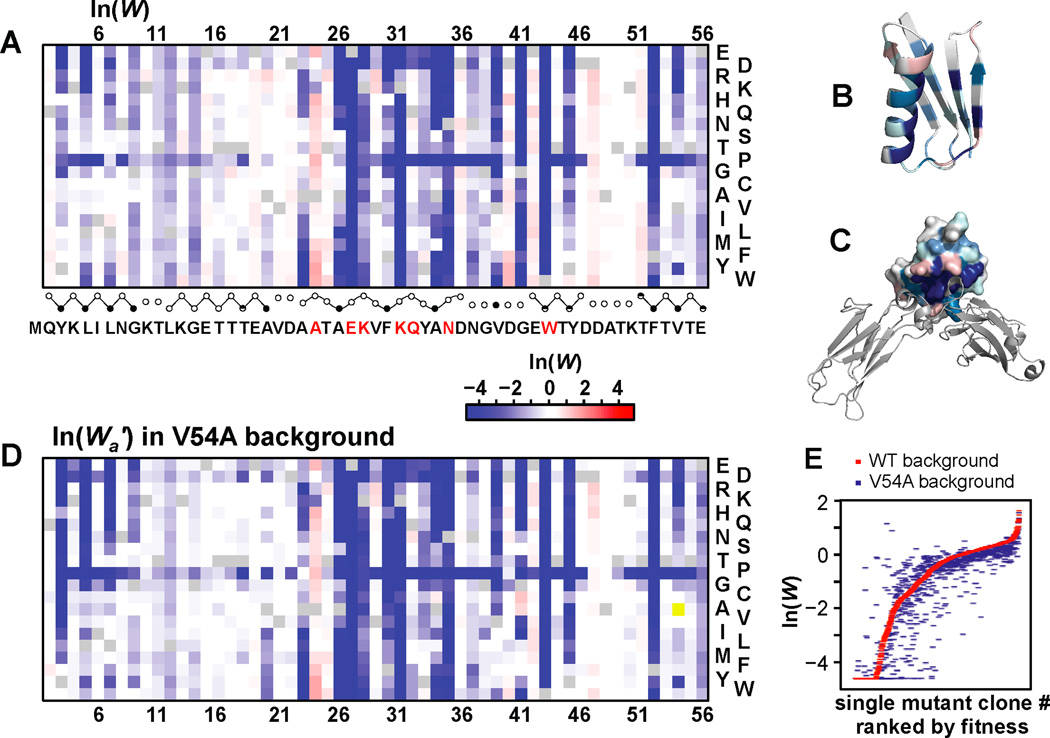
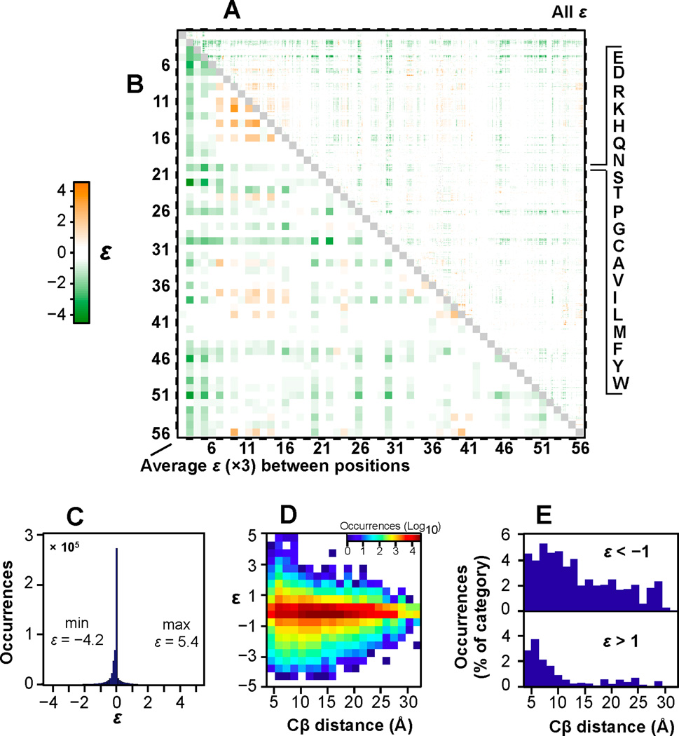
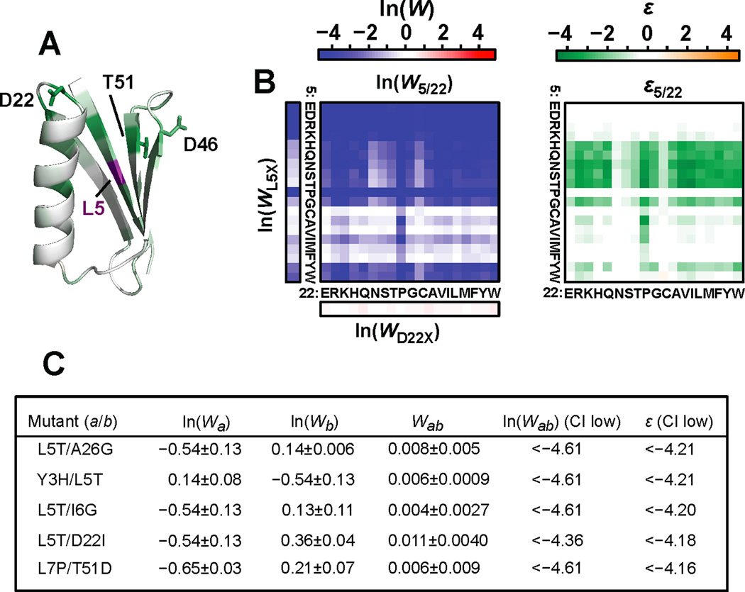
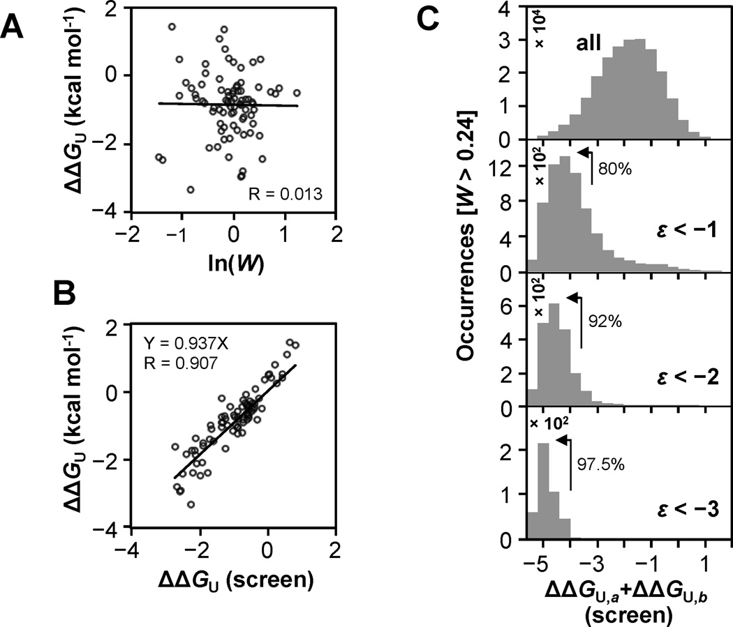
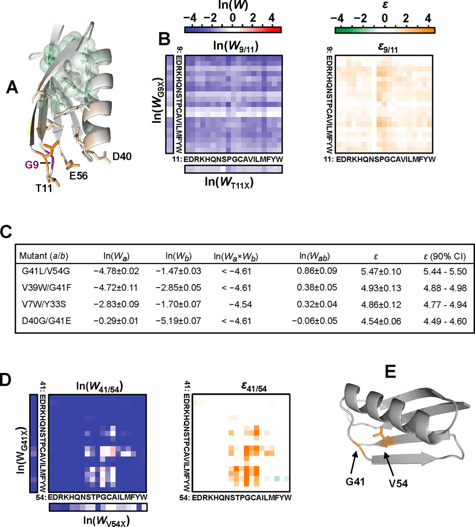
Results: To answer this question, we quantified the effects of all single mutations and double mutations between all positions in the IgG-binding domain of protein G (GB1). By observing the first two steps of all possible evolutionary pathways using this fitness profile, we were able to characterize the extent and magnitude of pairwise epistasis throughout an entire protein molecule. Furthermore, we developed a novel approach to quantitatively determine the effects of single mutations on structural stability (ΔΔGU). This enabled determination of the importance of stability effects in functional epistasis.

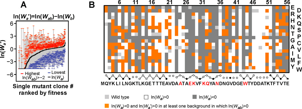
Conclusions: Our results illustrate common biophysical mechanisms for occurrences of positive and negative epistasis. Our results show pervasive positive epistasis within a conformationally dynamic network of residues. The stability analysis shows that significant negative epistasis, which is more common than positive epistasis, mostly occurs between combinations of destabilizing mutations. Furthermore, we show that although significant positive epistasis is rare, many deleterious mutations are beneficial in at least one alternative mutational background. The distribution of conditionally beneficial mutations throughout the domain demonstrates that the functional portion of sequence space can be significantly expanded by epistasis.


Figures

Figure 1
Figure 1. mRNA display fitness profile scheme
(A) A DNA library encoding all single and double mutants in GB1 was created (See also Figure S1). mRNA display was used to profile the relative binding efficiency of all variants. After a single generation of affinity enrichment, the relative fitness (W) for each variant was determined from the change in sequence frequency as identified by Illumina sequencing. (B) The binding fitness (red dots) of each single mutant and (C) all high-confidence double mutants (see Figure S1E). The gray dots represent the contribution to W from background binding to beads which was determined from a control lacking IgG-FC. (D) Thirteen clones were constructed and expressed in vitro with a 35S-Methionine label for comparison to fitness determined by the screen. Binding efficiency (see also Figure S1D) was used to estimate relative affinity (see the Experimental Procedures). Error bars representing standard deviation from the triplicated screen are shown (X-axis) and from the pull-down when performed in triplicate (Y-axis). (E) Correlation of Δln(KA) from the screen to Δln(KA) for 10 variants reported in the literature (see Table S3).
Figure 2
Figure 2. Affinity profile validation and fitness maps
(A) A heat-map depicting fitness of all single mutants. Residues previously determined to interact with IgG-FC [21] are highlighted in red. The fraction side-chain solvent accessibilities (closed<0.1, partial>0.1, open>0.4 circles) are depicted below. Circles are connected by straight or curved lines to delineate β-strands and the α-helix, respectively. (B) Average ln(W) plotted on GB1 (PDB 1PGA) [54] and (C) the complex between protein G domain C2 (space filled) and IgG-FC (cartoon) (PDB 1FCC) [21]. (D) A heat-map depicting fitness of all single mutants in the background of V54A. (E) Comparison of the fitness profile to fitness effects in the background of V54A. See also Figure S2.
Figure 3
Figure 3. Pairwise epistasis map throughout GB1
(A) A heat-map depicting epistasis for 517,278 double mutants (96.5% of all possible). The amino acid order is listed top to bottom and is the same left to right. Each of the 1485 pairs display 19×19 sequence variants. (B) Average ε for all sequence combinations at each positional combination multiplied by a factor of 3 in order to match the range of the color bar. (C) Histogram showing extent of epistasis (increments of 0.1). (D) Two dimensional histogram relating ε to Cβ distances in 1PGA [54]. (E) |ε|>1 as a percentage of total occurrences in each Cβ distance bin. See also Figure S3.
Figure 4
Figure 4. Positions display negative epistasis in general independent of amino acid combination
(A) Average ε between position 5 and all other positions in GB1 (1PGA) [54]. Distant surface residues that demonstrate negative epistasis are labeled. (B) Binding fitness and epistasis for all 361 combinations of substitutions for Leu5 and Asp22. Leu5 is a critical core residue that is sensitive to mutation while Asp22 is part of a helix stabilizing N-capping motif near the binding surface where substitutions are generally well tolerated for binding function. (C) The most significant values of negative epistasis are listed. Each pair includes mutations that are expected to destabilize the structure but alone do not unfold the protein or significantly disrupt affinity. A Poisson-based 90% confidence interval was used to generate an upper boundary for binding fitness thereby enabling a conservative estimate of epistasis. See also Figure S4.
Figure 5
Figure 5. Relationship between structural stability effects and epistasis
(A) Comparison of ln(W) to free energy of unfolding relative to wild type (ΔΔGU) reported in the literature. (B) The predicted thermodynamic stability of 82 single mutants compared to ΔΔGU values reported in the literature (See Table S4). ΔΔGU predicted by the screen are median values identified by estimating the change in fraction of unfolded protein in 5 destabilized mutant backgrounds. This analysis was limited to variants displaying W>0.24 (709 of 1045) as lower fitness values did not produce sufficient dynamic range to measure decreased structural stability. (C) Histograms of ΔΔGU,a+ΔΔGU,b showing how the distribution of predicted stability changes as the magnitude of negative epistasis increases. The percentage of each epistasis category resulting from combinations of significantly destabilized mutations (left of arrow) is listed. See also Figure S5.
Figure 6
Figure 6. Positions that display general positive epistasis independent of amino acid combination
(A) Average ε for all pairwise combinations with position 9. Glu56, which couples the two dynamic loops through H-bonds, is highlighted. (B) Epistasis and binding fitness for all 361 combinations of substitutions for G9 and T11 which are located within the highly dynamic β1–β2 loop. (C) The 20 most significant examples of positive epistasis include double mutants from 4 pairwise positional combinations. The double mutants displaying the largest value of positive epistasis per positional pair are listed. These combinations include neighboring residues within or at the edge of the conformationally dynamic region that overall demonstrates pervasive positive epistasis (see Figure 3A,B). For calculating epistasis, we limited expected fitness by ln(Wa×Wb)≥ln(0.01) to minimize spurious epistasis values for lethal or nearly lethal double mutants resulting from non-meaningful predicted fitness values below the background (~W=0.01). (D) Fitness and epistasis for all double mutants including positions 41 and 54. (E) Exchanging volume from core residue Val54 to Gly41 demonstrates the most extreme value of positive epistasis. See also Figure S6.
Figure 7
Figure 7. The fitness effects of many mutations change dramatically depending on the background in which they appear
(A) The range is bound by a blue dash for the lowest fitness in any of the 1026 possible alternate backgrounds, and by a red dash for the highest fitness. The highest fitness values are limited to double mutants displaying ln(Wab)>−2. (B) A map showing deleterious single mutants that are beneficial in at least one alternative mutational background even while limiting double mutant fitness greater than wild type (orange). See also Figure S7.

References

    1. Breen MS, Kemena C, Vlasov PK, Notredame C, Kondrashov FA. Epistasis as the primary factor in molecular evolution. Nature. 2012;490:535–538. - PubMed
    1. Kimura M. Recent development of the neutral theory viewed from the Wrightian tradition of theoretical population genetics. Proc. Natl. Acad. Sci. USA. 1991;88:5969–5973. - PMC - PubMed
    1. Weinreich DM, Watson RA, Chao L. Perspective: Sign epistasis and genetic constraint on evolutionary trajectories. Evolution. 2005;59:1165–1174. - PubMed
    1. Povolotskaya IS, Kondrashov FA. Sequence space and the ongoing expansion of the protein universe. Nature. 2010;465:922–926. - PubMed
    1. Harms MJ, Thornton JW. Evolutionary biochemistry: revealing the historical and physical causes of protein properties. Nat. Rev. Genet. 2013;14:559–571. - PMC - PubMed

Publication types

Substances