. 2015 Jan;163(1):113-9.
doi: 10.1016/j.imlet.2014.10.020. Epub 2014 Oct 23.

TCR and CD28 activate the transcription factor NF-κB in T-cells via distinct adaptor signaling complexes

Affiliations

TCR and CD28 activate the transcription factor NF-κB in T-cells via distinct adaptor signaling complexes

Youg Raj Thaker et al. Immunol Lett. 2015 Jan.

Abstract

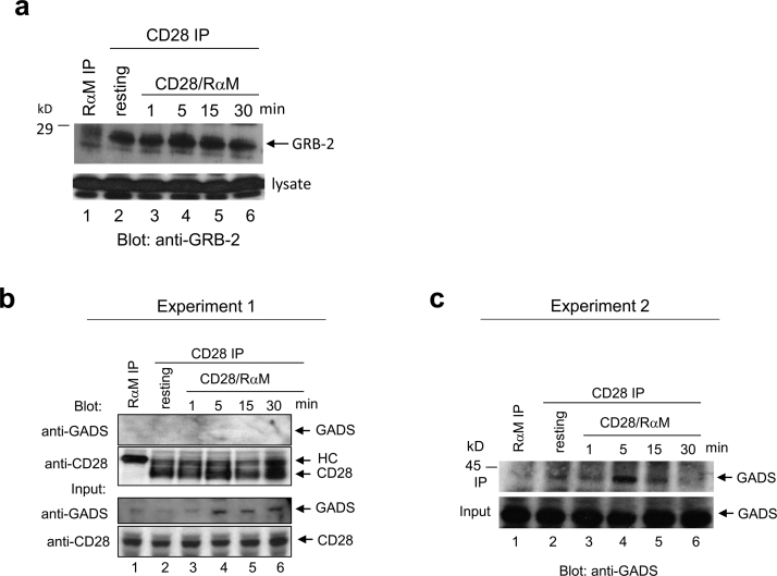
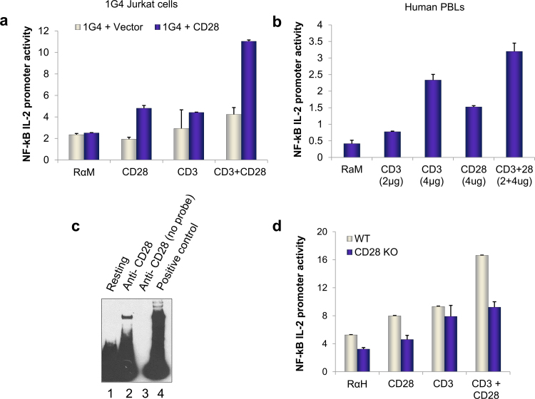
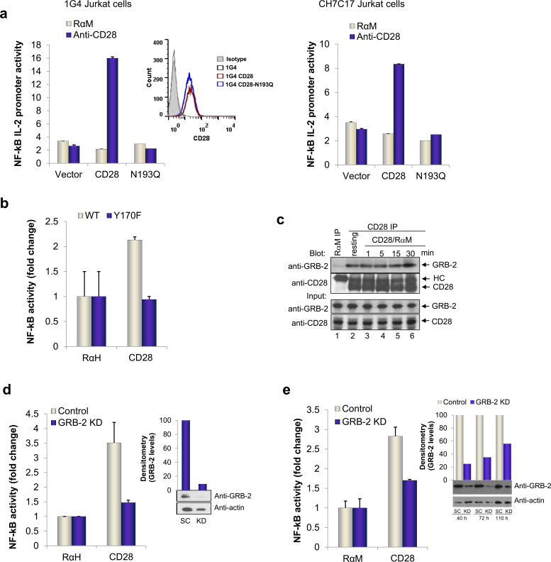
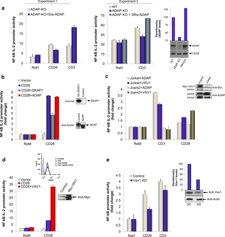
The transcription factor NF-κB is needed for the induction of inflammatory responses in T-cells. Whether its activation by the antigen-receptor and CD28 is mediated by the same or different intracellular signaling pathways has been unclear. Here, using T-cells from various knock-out (Cd28(-/-), adap(-/-)) and knock-in (i.e. Cd28 Y-170F) mice in conjunction with transfected Jurkat T-cells, we show that the TCR and CD28 use distinct pathways to activate NF-κB in T-cells. Anti-CD28 ligation alone activated NF-κB in primary and Jurkat T-cells as measured by NF-κB reporter and EMSA assays. Anti-CD28 also activated NF-κB normally in primary T-cells from adap(-/-) mice, while anti-CD3 stimulation required the adaptor ADAP. Over-expression of ADAP or its binding partner SKAP1 failed to enhance anti-CD28 activation of NF-κB, while ADAP greatly increased anti-CD3 induced NF-κB activity. By contrast, CD28 activation of NF-κB depended on GRB-2 binding to CD28 as seen in CD28 deficient Jurkat T-cells reconstituted with the CD28 YMN-FM mutant, and in primary T-cells from CD28 Y170F mutant knock-in mice. CD28 associated with GRB-2, and GRB-2 siRNA impaired CD28 NF-κB activation. GRB-2 binding partner and guanine nucleotide exchange factor, VAV1, greatly enhanced anti-CD28 driven activation of NF-κB. Further, unlike in the case of anti-CD28, NF-κB activation by anti-CD3 and its cooperation with ADAP was strictly dependent on LAT expression. Overall, we provide evidence that CD28 and the TCR complex regulate NF-κB via different signaling modules of GRB-2/VAV1 and LAT/ADAP pathways respectively.

Keywords: ADAP; Adaptors; GRB-2; NF-κB; VAV-1.


Figures

Supplementary Figure S1
Binding of Grb-2 and GADS to CD28. Jurkat T-cells, either left unstimulated (lane 2) or stimulated with anti-CD28/rabbit anti-mouse IgG for 1, 5, 15 and 30 min (lanes 3-6) were lysed, immunoprecipitated with anti-CD28 and blotted for Grb-2 (a) or GADS (b, c). Immunoprecipitation with rabbit anti-mouse IgG served as a negative control (lane 1). Input panels: blotting of cell lysates with Grb-2 or Gads served as a loading control.
Fig. 1
Fig. 1
CD28 and TCR uniquely regulated NF-κB. (a) 1G4 CD28 deficient Jurkat cells reconstituted with either empty plasmid (vector) or WT (CD28) untagged plasmid were used to measure NF-κB IL-2 promoter luciferase (firefly) activity in response to indicated ligating antibodies. Activity was measured by normalizing firefly luciferase counts to the background Renilla luciferase values, done in triplicates for each point as described in Materials and Methods section. All values are in relative luciferase units. (b) NF-κB activation in CD3+ human peripheral blood lymphocytes in response to engagement with indicated concentrations of CD3 (OKT3 clone) or CD28 (CD28.2 clone) alone or in combination. (c) Electromobility shift assay from nuclear fractions of C57BL/6 naïve T-cells as described in section 2. Briefly, a biotinylated NF-κB probe was used to detect DNA binding to nuclear fraction from unstimulated (lane 1) or anti-CD28 stimulated cells (lane 2). For specificity, anti-CD28 stimulated nuclear fraction was probed with unlabeled/cold NF-κB oligo (lane 3). Lane 4 is positive control (provided with kit). (d) NF-kB activation in primary T-cells isolated from WT or CD28 KO mice treated with control (RαH), anti-CD3 or anti-CD28 alone stimulating antibodies or in combination. Data in each sample is average of triplicates. All antibodies concentrations are in micrograms per milliliter of solution.
Fig. 2
Fig. 2
GRB-2 connects proximal signals to regulate CD28 driven NF-κB pathway. (a) Cd28−/− cells were reconstituted with empty vector, WT CD28 or N193Q mutant. FACS histogram shows surface expression of WT or N193Q CD28 by staining with FITC labeled antibodies to CD28. Right panel shows another representative experiment obtained from CH7C17, CD28 deficient cell line. NF-κB luciferase activity was measured after stimulation with indicated antibodies. (b) Y170F knock-in mutant primary T-cells were used in conjunction with wild type primary cells. Luciferase firefly units were normalized to background Renilla values. (c) Binding of Grb-2 to CD28. Jurkat T-cells, either left unstimulated (lane 2) or stimulated with anti-CD28/rabbit anti-mouse IgG for 1, 5, 15 and 30 min (lanes 3–6) were lysed, immunoprecipitated with anti-CD28 and blotted for Grb-2. Immunoprecipitation with rabbit anti-mouse IgG served as a negative control (lane 1). Input panels: blotting of cell lysates with Grb-2 (upper panel) or CD28 (lower panel) served as a loading control. siRNA knock-down of GRB-2 in primary cells (d) and Jurkat cells (e) and its effects on NF-κB pathway after 48 and 72 h of knock-down respectively. Efficiency of knock-down was assessed by western blotting as shown in insets (SC: scrambled and KD: knock-down) and quantified by normalizing to endogenous actin levels (inset column chart). All siRNA were purchased from Dharmacon (Thermo Scientific).
Fig. 3
Fig. 3
Normal CD28 but defective TCR mediated NF-κB pathway in ADAP KO primary and LAT deficient Jurkat cells. (a) CD3+ cells isolated from spleens of adap−/− (silver bars) or wild type (blue bars) mice were used for NF-κB IL-2 promoter assay by transfecting with reporter plasmids followed by stimulation with control, anti-CD28 or anti-CD3 antibodies. Right panel, NF-κB activity in adap−/− (blue bars) compared to controls wild type (silver bars) or adap−/− T-cells transfected with exogenous ADAP (green bars). Efficiency of transfection was assessed by western blotting and normalized to endogenous CD28 levels as shown by column chart (inset). Each point is average of triplicate with SD. (b) NF-κB activity measured in CD28 reconstituted Jurkat 1G4 cells transfected with adapters SKAP1 or ADAP (Fyb) or control plasmids as shown. NF-κB activity was measured as above. (c) NF-κB reporter activity in LAT deficient (Jcam2) cells, either transfected with VAV1 (green bars) or ADAP (silver bars) when engaged with control (RaM), anti-CD28 or anti-CD3 antibodies. Wild type (Jurkat) transfected with respective plasmids were used as control. All values are relative luciferase units and average of triplicates with standard deviations. (d) Reconstitution of Cd28−/− Jurkat cells with VAV1 or respective controls. NF-κB activation in response to CD28 was measured as above. (e) siRNA knock-down of VAV1 in primary T-cells for 48 h. NF-κB activity was measured from reporter plasmids when cells were stimulated for 6 h with control (RαH), anti-CD28 or anti-CD3 antibodies. Knock-down efficiency was detected by western blotting of endogenous proteins and quantified by densitometric analysis and shown as actin normalized values in column chart (inset). (For interpretation of the references to color in this figure legend, the reader is referred to the web version of this article.)

References

    1. Rudd C.E. Adaptors and molecular scaffolds in immune cell signaling. Cell. 1999;96:5–8. - PubMed
    1. Samelson L.E. Signal transduction mediated by the T cell antigen receptor: the role of adapter proteins. Annu Rev Immunol. 2002;20:371–394. - PubMed
    1. Weiss A. TCR signal transduction: opening the black box. J Immunol. 2009;183:4821–4827. - PubMed
    1. Dustin M.L., Shaw A.S. Costimulation: building an immunological synapse. Science. 1999;283:649–650. - PubMed
    1. June C.H., Bluestone J.A., Nadler L.M., Thompson C.B. The B7 and CD28 receptor families. Immunol Today. 1994;15:321–331. - PubMed

Publication types