. 2014 Nov 20;9(4):1256-64.
doi: 10.1016/j.celrep.2014.10.042. Epub 2014 Nov 13.

An active role for the ribosome in determining the fate of oxidized mRNA

Affiliations

An active role for the ribosome in determining the fate of oxidized mRNA

Carrie L Simms et al. Cell Rep. .

Abstract

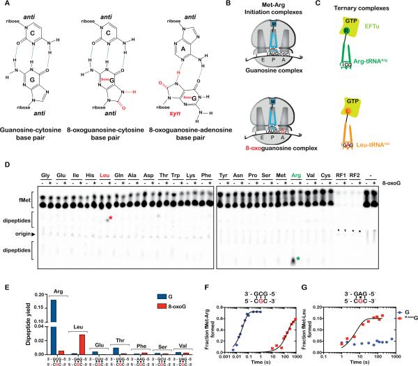
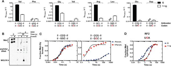
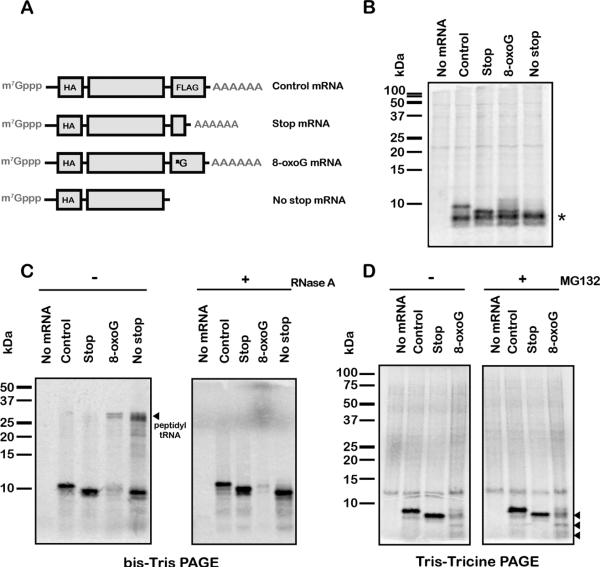
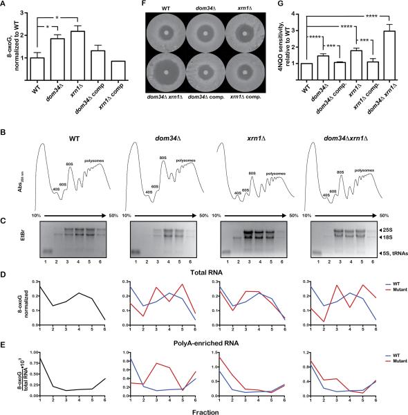
Chemical damage to RNA affects its functional properties and thus may pose a significant hurdle to the translational apparatus; however, the effects of damaged mRNA on the speed and accuracy of the decoding process and their interplay with quality-control processes are not known. Here, we systematically explore the effects of oxidative damage on the decoding process using a well-defined bacterial in vitro translation system. We find that the oxidative lesion 8-oxoguanosine (8-oxoG) reduces the rate of peptide-bond formation by more than three orders of magnitude independent of its position within the codon. Interestingly, 8-oxoG had little effect on the fidelity of the selection process, suggesting that the modification stalls the translational machinery. Consistent with these findings, 8-oxoG mRNAs were observed to accumulate and associate with polyribosomes in yeast strains in which no-go decay is compromised. Our data provide compelling evidence that mRNA-surveillance mechanisms have evolved to cope with damaged mRNA.


Figures

Figure 1
Figure 1. 8-oxoG is detrimental to the decoding process
(A) Chemical structures of G:C, 8-oxoG:C and 8oxoG:A base pairs. 8-oxoG adopts a syn conformation allowing it to base pair with adenosine. (B) Schematic representation of guanosine and 8-oxoguanosine initiation complexes encoding for the dipeptide Met-Arg. Both complexes carry the initiator fMet-tRNAfMet in the P site; the G complex displays a CGC codon in the A site, whereas the 8-oxoG complex displays a C8-oxoGC codon in the A site. (C) Schematic representation of the cognate Arg-tRNAArg and near-cognate Leu-tRNALeu (G:A mismatch at the second position) ternary complexes. (D) Phosphorimager scan of an electrophoretic TLC showing the reactivities of the initiation complexes with the indicated ternary complexes and release factors. Green asterisk represents the cognate dipeptide, whereas the red one represents the near-cognate dipeptide resulting from a G:A mismatch. (E) Quantification of the dipeptide yield in (D); the predicted codon-anticodon interaction is shown below the x-axis and the corresponding dipeptide is shown above the bars. (F, G) Time-courses of peptide-bond formation between the initiation complexes and the cognate Arg-tRNAArg and near-cognate Leu-tRNALeu ternary complexes, respectively. See also Figure S1, S2.
Figure 2
Figure 2. 8-oxoG inhibits peptide-bond formation
(A) Observed rates for peptidyl transfer (kPT) measured on native (GUU, GGC, CGC, GAG) and 8-oxoG complexes (8-oxoGUU, G8-oxoGC, C8-oxoGC, GA8-oxoG) with cognate - left two bars in each graph - and near-cognate (A:G mismatches at the 8-oxoG position) – right two bars in each graph tRNAs. The codon-anticodon interaction is shown below the x-axis and the corresponding dipeptide is shown above the bars. Clear bars represent rates observed with native complexes; black ones represent those observed with 8-oxoG complexes. Reactions were carried out at least in duplicate ± SEM. (B) Phosphorimager scan of an electrophoretic TLC of reactions between native complex (GGC) or 8-oxoG complex (G8-oxoGC), which encode MGLYK peptide, and the full complement of aa-tRNAs, elongation and release factors (PURE system, NEB). Reactions were allowed to proceed for 30 seconds before addition of 100mM KOH. (C) Time-courses of peptide-bond formation between the native GGC complex and Gly-tRNAGly (left panel) or oxidized G8oxoGC complex and Gly-tRNAGly (right panel) in the absence (blue circles) and presence of paromomycin (red squares). (D) Time-courses of RF2-mediated release on native UGA (blue circles) and U8-oxoGA (red squares) initiation complexes.
Figure 3
Figure 3. 8-oxoG stalls translation in cell extracts
(A) Schematic of the mRNA reporters used in eukaryotic extracts. The full-length mRNA encodes a peptide that has a molecular weight of ~9 kDa, whereas the stop mRNA, which has a stop codon at the position of the 8-oxoG codon in the 8-oxoG mRNA, encodes a peptide that has a molecular weight of ~8.4 kDa. Reporters used in S30 reactions were identical minus the cap and polyadenylation. (B) Autoradiograph of a bis-Tris gel of translation assays using bacterial S30 extracts. The 8-oxoG transcript yields a peptide of a size similar to the stop reporter. * indicates non-specific band. (C) Autoradiograph of bis-Tris and Tris-Tricine gels of in vitro translation assays in wheat germ extracts. Proteins were labeled by the addition of [35S]-Methionine to the reactions. The 8-oxoG mRNA and no-stop mRNA both accumulate peptidyl-tRNA, visible on the bis-Tris gel, which disappear upon the addition of RNase A. Reactions separated on Tris-Tricine were incubated with and without MG132. The 8-oxoG mRNA produces truncated protein products (marked by arrows); the largest of these has a size similar to that observed in the presence of stop mRNA. See also Figure S3.
Figure 4
Figure 4. 8-oxoG mRNA is stabilized in yeast in the absence of no-go-decay factors and associates with polysomes in the absence of Dom34
(A) Bar graph showing the levels of 8-oxoG, relative to WT, in polyA-enriched RNAs isolated from the indicated yeast strains. The values shown are the means of at least three measurements ±SEM. * p<0.05 (B) Polysomes profiles of the indicated strains. (C) Denaturing agarose electrophoresis of the RNA samples isolated from the different fractions in the sucrose gradient. (D, E) Distribution of 8-oxoG levels across the gradients in total RNA and polyA-enriched RNA samples, respectively. Levels of 8-oxoG in (D) were normalized to the total amount of 8-oxoG across the gradient, whereas in (E) they were divided by the amount of polyA-enriched RNA. (F) Representative images of zone of inhibition assays on the indicated yeast strains using 4-nitroquinoline-1-oxide (4NQO). (G) Quantification of 4NQO sensitivity for the indicated strains, relative to WT. Data are mean ±SEM, from results of at least three biological replicates. *** p<0.001, **** p<0.0001 See also Figure S4.

References

    1. Barciszewski J, Barciszewska MZ, Siboska G, Rattan SI, Clark BF. Some unusual nucleic acid bases are products of hydroxyl radical oxidation of DNA and RNA. Mol. Biol. Rep. 1999;26:231–238. - PubMed
    1. Carter AP, Clemons WM, Brodersen DE, Morgan-Warren RJ, Wimberly BT, Ramakrishnan V. Functional insights from the structure of the 30S ribosomal subunit and its interactions with antibiotics. Nature. 2000;407:340–348. - PubMed
    1. Doma MK, Parker R. Endonucleolytic cleavage of eukaryotic mRNAs with stalls in translation elongation. Nature. 2006;440:561–564. - PMC - PubMed
    1. Fernandez IS, Ng CL, Kelley AC, Wu G, Yu YT, Ramakrishnan V. Unusual base pairing during the decoding of a stop codon by the ribosome. Nature. 2013;500:107–110. - PMC - PubMed
    1. Finkel T, Holbrook NJ. Oxidants, oxidative stress and the biology of ageing. Nature. 2000;408:239–247. - PubMed

Publication types

LinkOut - more resources