. 2014 Nov 20;9(4):1333-48.
doi: 10.1016/j.celrep.2014.10.039. Epub 2014 Nov 13.

Regulation of Stat5 by FAK and PAK1 in Oncogenic FLT3- and KIT-Driven Leukemogenesis

Affiliations

Regulation of Stat5 by FAK and PAK1 in Oncogenic FLT3- and KIT-Driven Leukemogenesis

Anindya Chatterjee et al. Cell Rep. .

Abstract

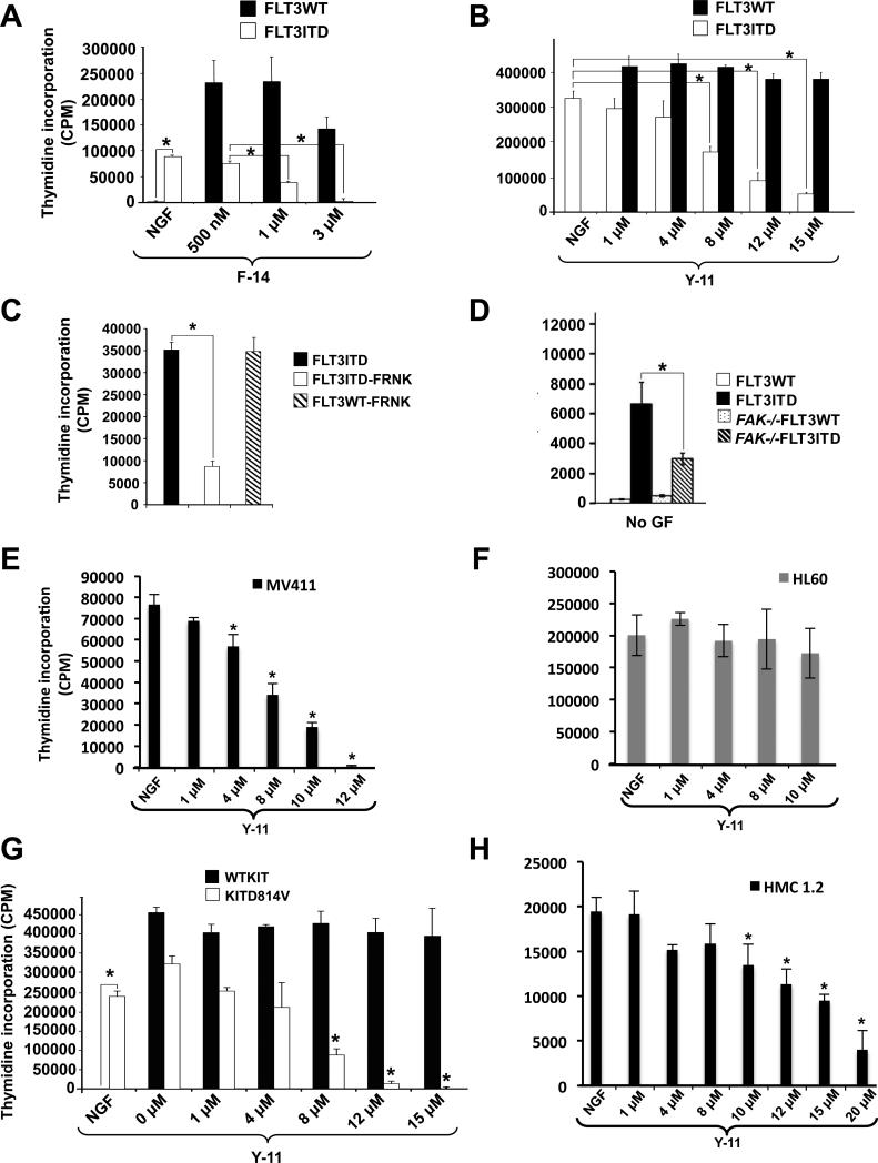
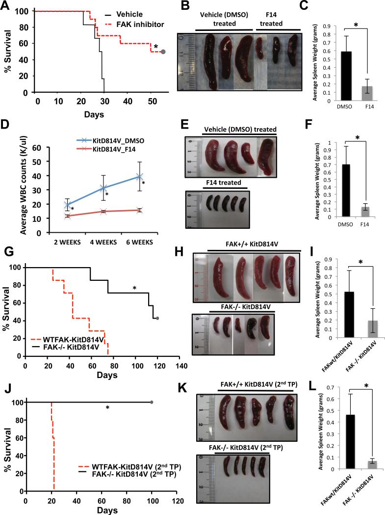
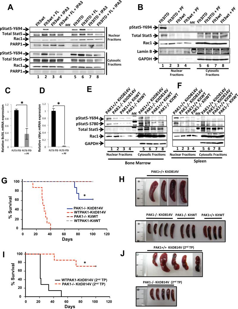
Oncogenic mutations of FLT3 and KIT receptors are associated with poor survival in patients with acute myeloid leukemia (AML) and myeloproliferative neoplasms (MPNs), and currently available drugs are largely ineffective. Although Stat5 has been implicated in regulating several myeloid and lymphoid malignancies, how precisely Stat5 regulates leukemogenesis, including its nuclear translocation to induce gene transcription, is poorly understood. In leukemic cells, we show constitutive activation of focal adhesion kinase (FAK) whose inhibition represses leukemogenesis. Downstream of FAK, activation of Rac1 is regulated by RacGEF Tiam1, whose inhibition prolongs the survival of leukemic mice. Inhibition of the Rac1 effector PAK1 prolongs the survival of leukemic mice in part by inhibiting the nuclear translocation of Stat5. These results reveal a leukemic pathway involving FAK/Tiam1/Rac1/PAK1 and demonstrate an essential role for these signaling molecules in regulating the nuclear translocation of Stat5 in leukemogenesis.


Figures

Fig.1
Fig.1. FAK is constitutively phosphorylated in FLT3 and activating KITD814V oncogene bearing cells
(A) Serum starved 32D cells expressing FLT3ITD and FLT3WT were treated with DMSO (lanes 1, 5); F-14 (lanes 2, 6); IL-3 (lanes 3, 7) or F-14 followed by IL-3 (lanes 4, 8). An equal amount of protein was subjected to western blot analysis and probed with phospho-FAK (Y397) antibody (n=3). ‘MK’ denotes lane with protein ladder. (B) 32D cells expressing KITWT or KITD814V were treated with F-14 (n=2) and analyzed as described in (A). (C) 32D cells expressing FLT3WT were treated with F-14 (lane 2) or stimulated with FLT3 ligand (FL) (lane 3), and analyzed as described in (A). (D) 32D cells expressing FLT3WT or FLT3ITD were treated with FLT3ITD inhibitor AC220 and analyzed for activated FLT3 (pY589/591). (E) 32D cells expressing FLT3ITD were treated with F-14 (lane 2) or with AC220 (lane 3) (n=2) and analyzed as above. (F) Lysates from primary FAK−/− deficient or WT BM cells expressing KITD814V or empty vector were analyzed for activated FAK. (G) FLT3ITD+ve AML patient sample was analyzed for activated FAK (n=2). (H) 32D cells expressing FLT3ITD were treated with Y-11 and subjected to flow cytometry analysis. The percentage of cells show activated FAK under basal conditions in FLT3WT (i), FLT3ITD vehicle treated cells (ii), and FLT3ITD treated with Y-11 (iii). n=2.
Fig.2
Fig.2. Inhibition of FAK suppresses the constitutive growth of oncogenic FLT3 and KIT bearing cells
(A) BaF3 or (B) 32D cells expressing FLT3WT or FLT3ITD were cultured for 48 hours in the presence or absence of F-14 or Y-11 in replicates of four and subjected to a thymidine incorporation assay. (C) BaF3 cells co-expressing FRNK and either FLT3ITD or FLT3WT were subjected to thymidine incorporation assay as in (A) & (B). (D) WT or FAK−/− BM cells expressing FLT3ITD or FLT3WT were subjected to proliferation assay in the absence of growth factors as described in (A) & (B). (E) MV4-11 cells expressing endogenous levels of FLT3ITD or (F) HL60 cells harboring FLT3WT were subjected to thymidine incorporation assay in presence of Y-11. (G) 32D cells expressing KITD814V or WTKIT and (H) HMC1.2 human leukemic cells line harboring KIT (D816V + G560V) mutations were cultured in the absence or presence of Y-11, and subjected to thymidine incorporation assay. Thymidine incorporation is depicted on y-axis as mean ± SD, *p<0.05. NGF/ No GF= cells grown in presence of no growth factors/cytokines. Data are representative of at least 3 independent experiments.
Fig.3
Fig.3. AC220 resistant FLT3 mutations, primary AML FLT3ITD+ cells or KITD816V+ SM cells are sensitive to FAK inhibition
(A) BaF3 cells bearing FLT3ITD or FLT3 receptors with acquired AC220 resistant mutations in the kinase domain (D835Y, F691L and D835V) were subjected to proliferation assay as described in Fig 2, *p<0.05. (B) Primary AML patient cells positive for FLT3ITD mutation (AML#1-4) or (C) primary KITD816V(+) or KITD816V(-) SM cells were treated with indicated concentrations of F-14 or Y-11. After 48 hours proliferation assay was performed. Bars denote mean ± SD, *p<0.05.
Fig.4
Fig.4. Rac1 is a downstream effector of FAK in FLT3ITD bearing oncogenic pathway
(A) BaF3 (lane 1) or 32D (lane 2) cells expressing the FLT3WT receptor or FLT3ITD both alone or in combination with FRNK were starved and subjected to a Rac activation assay. These cells were either vehicle treated alone (upper panel [CT]) or with F-14 (lower panel) (n=2). (B) MV4-11 cells expressing endogenous FLT3ITD, and (C) AML patient FLT3ITD+ cells were subjected to Rac activation assay as described in (A). (D) FLT3ITD bearing BM cells in the setting of FAK deficiency were subjected to Rac activation assay as in (A) (n=2). (E) 32D FLT3ITD cells expressing shRNA's targeting FAK (lane 2, 3) and control shRNA (lane 1) were subjected to Rac1 activation assay as described in (A).
Fig.5
Fig.5. FAK regulates the translocation of active Stat5 and Rac1 to the nucleus in FLT3ITD and KITD816V expressing cells
(A) 32D cells expressing FLT3ITD or FLT3WT were serum starved and subjected to fractionation assays, and nuclear and cytosolic fractions analyzed for levels of active Stat5 (pY694), total Stat5 and Rac1, and (B) 32D cells expressing FLT3ITD were subjected to fractionation assays after treatment with DMSO control, F-14, FLT3 ligand (FL) or with F-14 followed by FL (n=3). (C) HMC1.2 cells bearing KITD816V+G560V mutations, (D) MV4-11 and HL60 cells derived from leukemia patients harboring endogenous FLT3ITD and FLT3WT mutations were subjected to fractionation assay in presence of F-14 or Y-11 as in (A). Fractionation assays were performed in BM cells harvested from primary transplanted mice cohorts transplanted with KITD814V in a wild type FAK (FAK+/+) or FAK deficient (FAK−/−) background (n=2) (E). Fractionation assay from BM cells harvested from F-14 or DMSO (vehicle) treated primary transplant mice cohorts (F). The level of Stat5 phosphorylation/expression and Rac1 expression in the nuclear and cytosolic fractions is indicated. Expression of GAPDH was used as an indicator of cytosolic marker and loading control. ‘MK’ denotes lane with protein ladder. qRT-PCR analysis of relative mRNA expression levels of Stat5 responsive genes c-Myc (G) and BclXL (H) in FLT3ITD cells treated with F-14 or vehicle (DMSO) (n=2), *p<0.05.
Fig.6
Fig.6. Downstream of FAK, Tiam1 regulates the activation of Rac1 and subsequent translocation of active Stat5 to the nuclear compartment to develop leukemia in mice
(A) Nuclear fractions from F-14 treated FLT3ITD and FLT3WT bearing cells were subjected to Rac1 immunoprecipitation assay to assess the level of Rac1 binding to active Stat5. Level of active Stat5 (pY694), total Stat5 and Rac1 were analyzed. (B) Active Rac1 fractions from WT and FAK−/− deficient BM cells expressing FLT3ITD was determined, along with levels of active and total Stat5. Total Rac1 levels are shown in the lowermost panel (n=2). (C) Fractionation assay was performed using WT and Rac1−/− BM cells expressing FLT3ITD or FLT3WT receptors. Nuclear and cytosolic fractions were analyzed as described above. GAPDH was used as a loading control and cytosolic marker (n=3). ‘MK’ denotes lane with protein ladder. (D) 32D FLT3ITD and FLT3WT cells were treated with or without F-14 and subjected to Tiam1 activation assay (n=2). (E) 32D FLT3ITD and FLT3WT cells were subjected to Tiam1 IP n presence or absence of F-14. Samples were analyzed for amount of Rac1 binding Tiam1 (IP:Tiam1 panels). Lower two panels (Lysate (input)) depict the total protein levels of Rac1 and Tiam1 (n=2). (F) 32D cells co-expressing FLT3ITD and Tiam1 shRNA or scrambled shRNA were subjected to cellular fractionation assay and nuclear and cytosolic fractions were analyzed for the levels of active Stat5 (pY694), total Stat5 and Rac1, and nuclear marker/loading control PARP-1. (G) 32D cells co-expressing FLT3ITD and Tiam1 shRNA's (lanes 2-4) or scrambled shRNA (lane 1) were subjected to active Rac1 pull-down assay. The amount of active and total Rac1 are shown in the upper and lower panels, respectively. (H) Kaplan-Meier survival curve of mice transplanted with 32D cells co-expressing FLT3ITD and Tiam1 shRNA (n=5) or scrambled shRNA (n=5), (*p<0.01).
Fig.7
Fig.7. In vivo inhibition of FAK delays the development of MPN in mice transplanted with FLT3ITD and KITD814V bearing cells
(A) C3H/HeJ mice were transplanted with 32D cells bearing FLT3ITD, and treated with 20 mg/kg body weight F-14 for 28 days. Kaplan-Meier survival analysis of vehicle (n=14) vs. F-14 (n=15) treated mice showed significant increase in overall survival (*p<0.02), and significant reduction of spleen size and weight (B,C) in F-14 treated mice as compared to vehicle (DMSO) control treated mice. (D) BoyJ mice were irradiated and transplanted with 5-FU treated BM cells expressing KITD814V. Mice were randomly divided into 2 groups and treated with vehicle (DMSO) (n=7) or F-14 (n=7) for 3 weeks post-transplantation. Peripheral blood from mice was analyzed at intervals of 2, 4 and 6 weeks (D). After 6 weeks mice were harvested to determine spleen size (E) and weight (F). (G) 5-FU treated BM cells from WTFAK or FAK−/− mice expressing KITD814V were transplanted into lethally irradiated C57BL/6 mice. Kaplan-Meier survival analysis of FAK+/+ KITD814V (n=9) vs. FAK−/− KITD814V (n=9) mice, spleen size (H) and weight (I) is shown (*p<0.002). (J) Secondary transplants were performed using BM from FAK+/+KITD814V and FAK−/−KITD814V primary recipients. Kaplan-Meier survival analysis of FAK+/+ KITD814V (n=5) vs. FAK−/− KITD814V (n=5) is shown (*p<0.003), and spleen size (K) and weight (L) (*p<0.05).
Fig.8
Fig.8. In vivo inhibition of PAK1 delays the onset of MPN in mice transplanted with FLT3ITD bearing cells and inhibits the growth of leukemic patient cells
(A) 32D cells expressing FLT3ITD or FLT3WT were starved of serum and treated with the PAK inhibitor IPA-3 (lanes 3,7) alone, with FLT3 ligand (FL) (lanes 2,6), or with IPA-3 followed by FL (lanes 4,8) as indicated, and subjected to cellular fractionation assay. Nuclear and cytosolic fractions were quantitated and equal lysates were loaded on a gel and probed with the indicated antibodies. Arrows indicate the activation/expression of the labeled molecules in nuclear as well as in cytosolic fractions of FLT3ITD and FLT3WT bearing cells. Expression of PARP-1 was used as an indicator of nuclear loading (n=2). (B) 32D FLT3ITD and FLT3WT were serum starved and treated with the PAK inhibitor PF-3758309 (PF) and analyzed as described in (A). qRT-PCR analysis of relative mRNA expression levels of Stat5 responsive genes BclXL (C) and c-Myc (D) in FLT3ITD cells treated with PAK inhibitor PF-3758309 (n=2), *p<0.05. Fractionation assays were performed in BM cells (E) and splenocytes (F). Mice cohorts transplanted with KITD814V in a WT PAK1 (PAK1+/+KIT814V) or PAK1 deficient (PAK1−/−KITD814V) cells (n=2). The level of phospho-Stat5, total Stat5 and Rac1 in the nuclear and cytosolic fractions is indicated. Expression of GAPDH was used as an indicator of cytosolic marker and loading control. ‘MK’ denotes lane with protein ladder. (G) Primary transplant were carried out using 5-FU treated BM cells from WTPAK1 or PAK1−/− mice transduced with KITD814V or KITWT, and transplanted into lethally irradiated C57BL/6 mice. Four groups of mice were used: WTPAK1 KITD814V (n=8), WTPAK1 KITWT (n=5), PAK1−/− KITD814V (n=8), and PAK1−/− KITDWT (n=5). Kaplan-Meier survival analysis of PAK1+/+ KITD814V vs PAK1−/− KITD814V, WTPAK1 KITWT, PAK1−/−KITWT mice showed significant overall survival (*p<0.0003, Hom-Sidak Method), and significant reduction of spleen size (H). (I) Secondary transplants were performed using BM cells from PAK1+/+KITD814V and PAK1−/−KITD814V mice and transplanted into irradiated C57BL/6 mice. Kaplan-Meier survival analysis of PAK1+/+ KITD814V (n=5) vs PAK1−/− KITD814V (n=5) mice showed significant overall survival (*p<0.001), and significant reduction of spleen size (J).

References

    1. Aksamitiene E, Achanta S, Kolch W, Kholodenko BN, Hoek JB, Kiyatkin A. Prolactin-stimulated activation of ERK1/2 mitogen-activated protein kinases is controlled by PI3-kinase/Rac/PAK signaling pathway in breast cancer cells. Cell Signal. 2011;23:1794–1805. - PMC - PubMed
    1. Barros P, Lam EW, Jordan P, Matos P. Rac1 signalling modulates a STAT5/BCL-6 transcriptional switch on cell-cycle-associated target gene promoters. Nucleic acids research. 2012;40:7776–7787. - PMC - PubMed
    1. Baumgartner C, Cerny-Reiterer S, Sonneck K, Mayerhofer M, Gleixner KV, Fritz R, Kerenyi M, Boudot C, Gouilleux F, Kornfeld JW, et al. Expression of activated STAT5 in neoplastic mast cells in systemic mastocytosis: subcellular distribution and role of the transforming oncoprotein KIT D816V. The American journal of pathology. 2009;175:2416–2429. - PMC - PubMed
    1. Beghini A, Ripamonti CB, Cairoli R, Cazzaniga G, Colapietro P, Elice F, Nadali G, Grillo G, Haas OA, Biondi A, et al. KIT activating mutations: incidence in adult and pediatric acute myeloid leukemia, and identification of an internal tandem duplication. Haematologica. 2004;89:920–925. - PubMed
    1. Benekli M, Baer MR, Baumann H, Wetzler M. Signal transducer and activator of transcription proteins in leukemias. Blood. 2003;101:2940–2954. - PubMed

Publication types

MeSH terms