. 2014 Nov 20;9(4):1402-1416.
doi: 10.1016/j.celrep.2014.10.028. Epub 2014 Nov 13.

FXR1P limits long-term memory, long-lasting synaptic potentiation, and de novo GluA2 translation

Affiliations

FXR1P limits long-term memory, long-lasting synaptic potentiation, and de novo GluA2 translation

Denise Cook et al. Cell Rep. .

Abstract

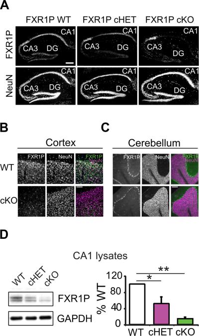
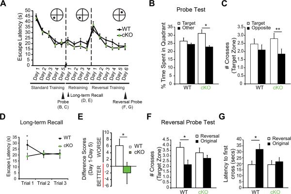
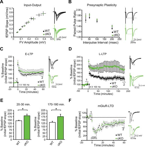
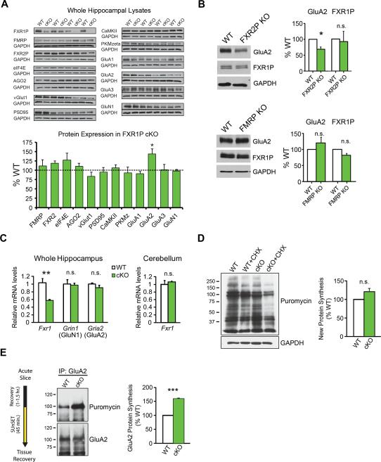
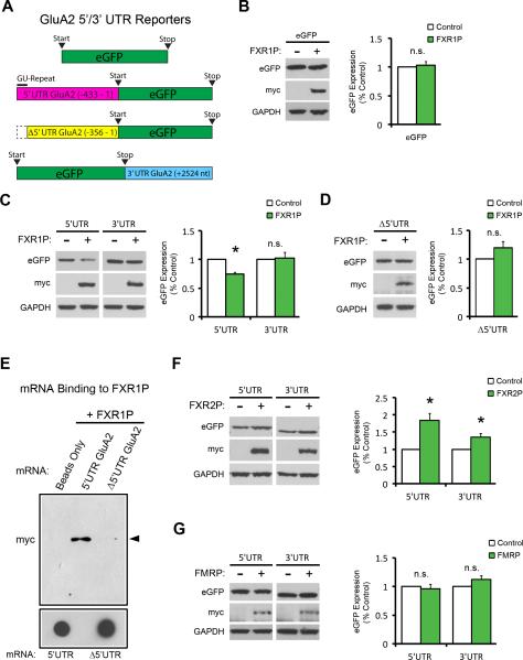
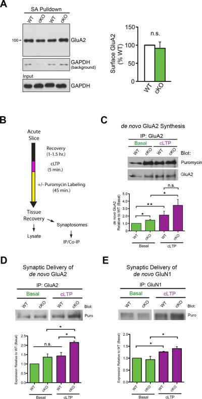
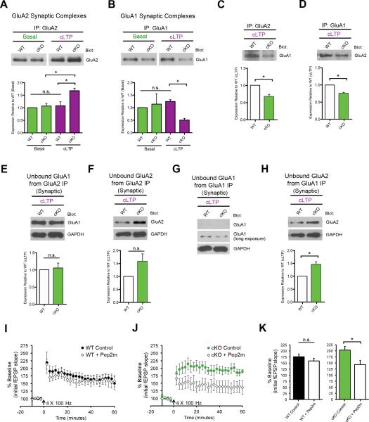
Translational control of mRNAs allows for rapid and selective changes in synaptic protein expression that are required for long-lasting plasticity and memory formation in the brain. Fragile X Related Protein 1 (FXR1P) is an RNA-binding protein that controls mRNA translation in nonneuronal cells and colocalizes with translational machinery in neurons. However, its neuronal mRNA targets and role in the brain are unknown. Here, we demonstrate that removal of FXR1P from the forebrain of postnatal mice selectively enhances long-term storage of spatial memories, hippocampal late-phase long-term potentiation (L-LTP), and de novo GluA2 synthesis. Furthermore, FXR1P binds specifically to the 5' UTR of GluA2 mRNA to repress translation and limit the amount of GluA2 that is incorporated at potentiated synapses. This study uncovers a mechanism for regulating long-lasting synaptic plasticity and spatial memory formation and reveals an unexpected divergent role of FXR1P among Fragile X proteins in brain plasticity.


Figures

Figure 1
Figure 1. Characterization of FXR1P cKO mice
A. Hippocampal sections taken from postnatal day 60 WT, cHET and cKO mice labeled for FXR1P and NeuN. FXR1P is lost from CA1 cells in cHET and cKO mice (n=3 mice/genotype). Scale bar=200μm. B, C. FXR1P is lost from cortical neurons (B), but not cerebellar Purkinje cells (C). Scale bar=100μm. D. Western blots of CA1 lysates from WT, cHET and cKO mice. FXR1P is reduced in the cHET and cKO mice (one-way ANOVA, F(2, 6) =16.66, p=0.004, Tukey HSD post-hoc, p<0.05; 3 mice/genotype from 3 litters). Error bars show standard error. **p≤0.01 *p≤0.05. See also Figure S1.
Figure 2
Figure 2. Loss of FXR1P enhances long-term memory and behavioral flexibility
A. cKO mice perform similarly to WT mice in the learning trials of Morris water maze training (two-way mixed ANOVA (Genotype, Days), main effect Days, F(4, 208) =46.52, p<0.001), retraining (two-way mixed ANOVA (Genotype, Days), p>0.05) and reversal training (two-way mixed ANOVA (Genotype, Days), main effect Days, F(5, 180) =24.73, p<0.001). B, C. cKO mice perform better than WT mice on the probe test 4 hours after the third trial on training day 5. cKO mice spend more time in the target quadrant vs. the average of all other quadrants (two-tailed paired t-test, t(27)=2.73, p=0.01), whereas WT mice do not (two-tailed paired t-test, t(27)=0.6784, p=0.50) (B). cKO mice cross the platform in the target zone more frequently than the platform in the opposite zone (two-tailed paired t-test, t(27)=2.79, p=0.01), whereas WT mice do not (two-tailed paired t-test, t(27)=0.79, p=0.44) (C). D, E. cKO mice have enhanced ability to recall long-term memories 9 days after the original 5 training days. cKO are faster at locating the hidden platform on the first trial of the first day of re-training (two-tailed unpaired t-test, t(33)=2.01, p=0.05) (D). cKO mice performed equally well on the first day of retraining (average of three trials) compared to their performance on the last day of the original learning phase (average of 3 trials), whereas WT mice are slower (two-tailed unpaired t-test, t(31)=2.19, p=0.04) (E). F, G. cKO mice do not show a preference towards the new platform location in the reversal probe test. WT mice cross the new platform location more times than the old platform location during the probe test (two-tailed paired t-test, t(19)=2.49, p=0.02), whereas cKO do not (two-tailed paired t-test, t(19)=0.92, p=0.37) (F). WT mice display a shorter latency to first cross for the new platform location vs. the old platform location (two-tailed paired t-test, t(19)=2.40, p=0.03), whereas cKO mice show equivalent latency to first cross of the reversal and original platform locations (two-tailed paired t-test, t(19)=0.57, p=0.57) (G). Error bars show standard error. *p≤0.05, **p≤0.01. See also Figure S2.
Figure 3
Figure 3. FXR1P cKO mice show a specific enhancement in L-LTP
A. Left Input-output curve showing synaptic responses upon increasing stimulation intensities for WT and cKO mice. Right Representative traces from WT and cKO mice. B. Left Paired-pulse facilitation (PPF) is normal in cKO mice (two-way mixed ANOVA, Genotype x Inter-stimulus interval, F(3,39) =0.44, p=0.73). Right Representative traces at a 50ms interval. C. Left E-LTP induced by a single train of high frequency stimulation (HFS: 1×100 Hz) is normal in cKO mice (n=8 mice, 9 slices, from 4 litters)(analysis at 50-60 minutes post-HFS, two-tailed unpaired t-test, t(15)=-0.38, p=0.71). Right Traces representing (1) 5 minutes of baseline immediately preceding HFS and (2) the period from 55-60 minutes post-LTP. D. Left Four trains of HFS delivered at 20s intervals (HFS: 4×100Hz) produce higher levels of potentiation in cKO animals (n=7 mice, 7 slices, from 5 litters) than WT animals. E. L-LTP, measured at 20-30 minutes and 170-180 minutes post-HFS, is greater in cKO (two-tailed unpaired t-tests, t(7)=2.65, p=0.03 and t(9)=2.24, p=0.05 respectively). Right Traces representing (1) 5 minutes of baseline immediately preceding HFS and (2) the period from 175-180 minutes post-LTP. F. Left mGluR-dependent LTD is unchanged in cKO mice (two-tailed, unpaired two-sample t-test, p=0.86). Right Traces representing (1) 5 minutes of baseline immediately preceding bath application of DHPG (100μM, 7-8 min) and (2) the period from 55-60 minutes post-DHPG administration. Error bars show standard error. Traces show an average of approximately 5-10 sweeps.
Figure 4
Figure 4. Enhanced GluA2 translation in FXR1P cKO mice
A. Top Analysis of Fragile X family proteins, molecules involved in protein synthesis, and synaptic proteins in WT and cKO mice (four pairs of animals). Most proteins are similarly expressed in both genotypes (two-tailed one-sample t-tests, p>0.05) with the exception of GluA2 (two-tailed one-sample t-test, p=0.02). Each blot was run 2-3X and values averaged. B. Top Blots of hippocampal lysates from WT and FXR2P KO mice showing levels of GluA2, FXR1P, and GAPDH. GluA2 is reduced (two-tailed, one-sample t-test, p=0.05; n=3 WT/3 FXR2P KO mice) and FXR1P is unchanged in FXR2P KO mice (two-tailed, one-sample t-test, p=0.26; n=3 WT/3 KO mice). Bottom Blots of hippocampal lysates from WT and FMRP KO mice showing levels of GluA2, FXR1P, and GAPDH. GluA2 and FXR1P are unchanged in FMRP KO mice (two-tailed, one-sample t-test, p>0.05; 3 WT/3 FMRP KO mice). C. qRT-PCR shows that Fxr1 mRNA levels are reduced in hippocampal lysates from FXR1P cKO mice (two-tailed, unpaired two-sample t-test, p=0.005; n=3 WT/4 cKO mice from 3 litters). The mRNA levels of Gria2 (GluA2) and Grin1 (GluN1) are unchanged (two-tailed, unpaired two-sample t-tests, p>0.05; n=3 WT/4 cKO mice). Fxr1 mRNA levels are unaltered in the cKO cerebellum (two-tailed, unpaired two-sample t-test, p>0.05; n=3 WT/3 cKO mice from 3 litters). D. Puromycin-labeled lysates from WT and cKO mice in the presence or absence of cycloheximide (CHX). No differences in overall puromycin labeling are detected between genotypes (p>0.05; 3 WT/3 cKO mice). E. IP for GluA2 shows increased puromycin-labeled GluA2 in slices from cKO mice, indicating enhanced GluA2 translation (p=0.002; 3 WT/3 cKO mice). Unless otherwise stated, statistical analyses were performed using two-tailed, one-sample t-tests. Error bars show standard error. ***p≤0.001, **p≤0.01, *p≤0.05, n.s.=not significant. See also Figures S3 and S4.
Figure 5
Figure 5. FXR1P represses translation through a GU-rich element in the GluA2 5’UTR
A. Constructs used in GluA2 reporter assay; eGFP, eGFP with the GluA2 5’UTR (-433 to 1), eGFP with the 5’UTR GU-rich element removed (Δ5’UTR (-356 to 1)), and eGFP with the GluA2 3’UTR (+2524 nt). B. FXR1P does not affect expression of eGFP alone (p=0.62). C. FXR1P represses expression from the GluA2 5’UTR-eGFP (p=0.004) but not the GluA2 eGFP-3’UTR construct (p=0.84). D. Deletion of the GU-rich element relieves FXR1P-mediated repression. E. Loss of the GU-rich element prevents binding of FXR1P to the 5’UTR of GluA2. F. FXR2P increases eGFP expression from constructs containing either the GluA2 5’UTR (p=0.02) or 3’UTR (p=0.05). G. FMRP does not regulate expression from either construct (p>0.05). Analyses were performed using two-tailed, one-sample t-tests. n=3-4 separate cultures and transfections. Error bars show standard error. * p≤0.05, n.s.=not significant. See also Figure S5.
Figure 6
Figure 6. FXR1P controls activity-dependent synaptic delivery of GluA2
A. Basal surface GluA2 levels are similar between genotypes (p=0.66; n=3 WT/3 cKO mice). Only low levels of GAPDH were biotinylated during the surface labeling process and purified with streptavidin (SA) beads (GAPDH background). B. Time course for cLTP experiments. C. cKO slices show increased basal de novo synthesis of GluA2 (p=0.04). Both WT and cKO slices increase GluA2 synthesis upon cLTP (WT: p=0.02 and cKO: p=0.05, two-tailed, unpaired two-sample t-test). D. cKO slices show increases in GluA2 synaptic delivery following cLTP (3 WT/3 cKO mice; p=0.01). E. GluN1 synaptic delivery is similar between genotypes (3 WT/3 cKO mice; p>0.05). Unless otherwise stated, statistical analyses were performed using two-tailed, one-sample t-tests. Error bars show standard error. **p≤0.01, *p≤0.05, n.s.=not significant. See also Figure S6.
Figure 7
Figure 7. FXR1P controls activity-dependent AMPAR composition
A. cKO slices show increases in GluA2-containing AMPARs at synapses following cLTP (3 WT/3 cKO mice; p=0.03). B. cKO slices show decreases in GluA1-containing AMPARs at synapses following cLTP (3 WT/3 cKO mice; p=0.002). C-D. cKO slices show decreases in association of GluA1 with GluA2 following cLTP in cKO slices (3 WT/3 cKO mice; p=0.04, p=0.01 respectively). E-H. Blotting unbound fractions from co-precipitations (C, D) show the amount of residual GluA1 and GluA2 molecules. I-J. cKO slices pre-incubated with myristoylated Pep2m show a significant reduction in potentiation at 20-30 minutes post L-LTP induction (WT (+/− pep2m): p=0.36, cKO (+/− pep2m): p=0.04, two-tailed unpaired t-tests). Unless otherwise stated statistical analyses were performed using two-tailed, one-sample t-tests *p≤0.05, n.s.=not significant. See also Figure S7.

References

    1. Ascano M, Mukherjee N, Bandaru P, Miller JB, Nusbaum JD, Corcoran DL, Langlois C, Munschauer M, Dewell S, Hafner M, et al. FMRP targets distinct mRNA sequence elements to regulate protein expression. Nature. 2012;492:382–386. - PMC - PubMed
    1. Bontekoe CJM, McIlwain KL, Nieuwenhuizen IM, Yuva-Paylor L. a, Nellis A, Willemsen R, Fang Z, Kirkpatrick L, Bakker CE, McAninch R, et al. Knockout mouse model for Fxr2: a model for mental retardation. Hum. Mol. Genet. 2002;11:487–498. - PubMed
    1. Cook D, Sanchez-Carbente M. del R., Lachance C, Radzioch D, Tremblay S, Khandjian EW, DesGroseillers L, Murai KK. Fragile X related protein 1 clusters with ribosomes and messenger RNAs at a subset of dendritic spines in the mouse hippocampus. PLoS One. 2011;6:e26120. - PMC - PubMed
    1. Cook D, Nuro E, Murai KK. Increasing our understanding of human cognition through the study of Fragile X Syndrome. Dev. Neurobiol. 2014;74:147–177. - PMC - PubMed
    1. Costa-Mattioli M, Gobert D, Harding H, Herdy B, Azzi M, Bruno M, Bidinosti M, Ben Mamou C, Marcinkiewicz E, Yoshida M, et al. Translational control of hippocampal synaptic plasticity and memory by the eIF2alpha kinase GCN2. Nature. 2005;436:1166–1173. - PMC - PubMed

Publication types