. 2015 Mar;62(3):581-9.
doi: 10.1016/j.jhep.2014.10.016. Epub 2014 Oct 18.

Phenotypic and functional analyses show stem cell-derived hepatocyte-like cells better mimic fetal rather than adult hepatocytes

Affiliations

Phenotypic and functional analyses show stem cell-derived hepatocyte-like cells better mimic fetal rather than adult hepatocytes

Melissa Baxter et al. J Hepatol. 2015 Mar.

Abstract

Background & aims: Hepatocyte-like cells (HLCs), differentiated from pluripotent stem cells by the use of soluble factors, can model human liver function and toxicity. However, at present HLC maturity and whether any deficit represents a true fetal state or aberrant differentiation is unclear and compounded by comparison to potentially deteriorated adult hepatocytes. Therefore, we generated HLCs from multiple lineages, using two different protocols, for direct comparison with fresh fetal and adult hepatocytes.

Methods: Protocols were developed for robust differentiation. Multiple transcript, protein and functional analyses compared HLCs to fresh human fetal and adult hepatocytes.

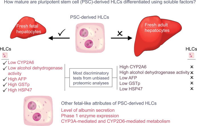
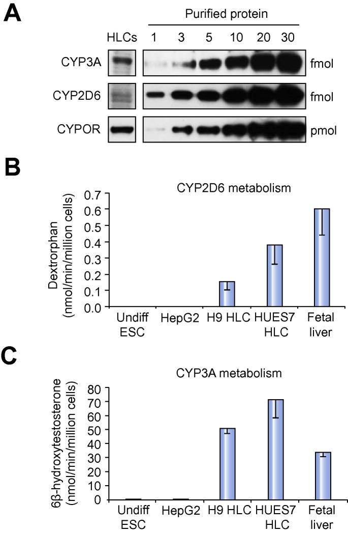
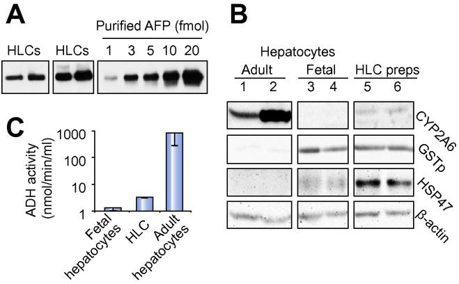
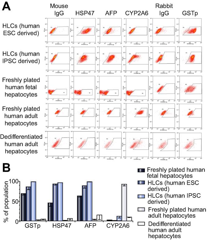
Results: HLCs were comparable to those of other laboratories by multiple parameters. Transcriptional changes during differentiation mimicked human embryogenesis and showed more similarity to pericentral than periportal hepatocytes. Unbiased proteomics demonstrated greater proximity to liver than 30 other human organs or tissues. However, by comparison to fresh material, HLC maturity was proven by transcript, protein and function to be fetal-like and short of the adult phenotype. The expression of 81% phase 1 enzymes in HLCs was significantly upregulated and half were statistically not different from fetal hepatocytes. HLCs secreted albumin and metabolized testosterone (CYP3A) and dextrorphan (CYP2D6) like fetal hepatocytes. In seven bespoke tests, devised by principal components analysis to distinguish fetal from adult hepatocytes, HLCs from two different source laboratories consistently demonstrated fetal characteristics.

Conclusions: HLCs from different sources are broadly comparable with unbiased proteomic evidence for faithful differentiation down the liver lineage. This current phenotype mimics human fetal rather than adult hepatocytes.

Keywords: Embryo; Hepatic; Hepatotoxicity; Human embryonic stem cell; Liver.


Figures

None
Graphical abstract
Fig. 1
Fig. 1
The three-stage differentiation protocol. RPMI, Roswell Park Memorial Institute; FBS, fetal bovine serum.
Fig. 2
Fig. 2
Differentiation of ESCs to hepatocyte-like cells. (A) Immunoblotting of ESCs differentiated towards definitive endoderm (DE). (B) Inclusion of Wnt3A for stage 1a improved detection of FOXA2, SOX17, and GATA4 by immunoblotting and immunofluorescence at day 4 (DE). Size bar = 25 μm. (C). Mean percentage of cells by count (± S.E.; numbers represented by the bar are shown below) that contained nuclear FOXA2, SOX17 or GATA4 either alone or in combination with NANOG at the end of stage 1. (D) Brightfield image of HLCs (H9 shown), compared to freshly plated human adult hepatocytes. Size bar = 150 μm. (E) Quantification by cell counting (mean ± S.E.) of dual immunofluorescence for albumin and α1-antitrypsin (AAT) in the five human ESC lines. Example image is for the H9 lineage counterstained with DAPI. Size bar = 50 μm. (F) Albumin secretion into the media (mean ± S.E. from >3 experiments) during differentiation compared to the secretion from equivalent numbers of freshly plated human fetal and adult hepatocytes. n.s., no significant difference between day 23 and day 30 for all lines (except HUES7 at day 30, p <0.05) and fetal hepatocytes. Values for all lines except HUES8 (n.s.) were significantly lower (p <0.05) than for adult hepatocytes.
Fig. 3
Fig. 3
Proteins upregulated in HLCs characterize liver more than other human organs and tissues. Heatmap of the 61 proteins significantly upregulated (>2-fold) in H9 HLCs compared to undifferentiated ESCs (Supplementary Table 2) analysed against gene expression experiments from a wide range of human organs and tissues, deposited in the EMBL/EBI Gene Expression Atlas (GEA). The numbers in individual red or blue boxes represent the number of experiments deposited in the GEA database where the gene, encoding that particular gene was up- (red) or downregulated (blue) in the relevant organ or tissue type.
Fig. 4
Fig. 4
Quantitative RT-PCR expression analysis of genes encoding phase 1 enzymes. (A–F) Genes encoding different classes of phase 1 enzymes. Gene expression in HLCs, quantified as fold difference over levels in the corresponding undifferentiated ESCs. Fold difference in fresh human adult hepatocytes (black numbers above the bars) and fresh human fetal hepatocytes (grey numbers above the bars) are relative to levels in undifferentiated H9 cells. For light blue (H9 HLCs) and dark blue (HUES7 HLCs) symbols next to bars: p <0.05, ∗∗p <0.02, p <0.01, p <0.005, ^p <0.001 compared to their parent undifferentiated ESCs. For symbols next to the black (adult) or grey (fetal) numbers, indicates no statistical difference from H9 HLCs; when encircled (for CYP1A1 and CYP1B1) the H9 HLC level was statistically higher than fetal or adult expression levels.
Fig. 5
Fig. 5
HLCs are metabolically similar to fetal hepatocytes. (A) Immunoblotting for CYP3A, CYP2D6 and CYPOR in HLCs. Equivalent numbers of human adult hepatocytes contained approximately 30 fmol of CYP3A and 20 fmol of CYP2D6. (B) CYP2D6 metabolism of dextromethorphan to dextrorphan. Conversion in fresh adult hepatocytes was 200 nmol/min/million cells. (C) CYP3A metabolism of testosterone to 6β-hydroxytestosterone. Conversion in fresh adult hepatocytes was 3300 nmol/min/million cells. Bar graphs in (B-C) show mean ± S.E. from >3 independent experiments.
Fig. 6
Fig. 6
Bespoke tests to distinguish hepatocyte maturity. (A–C) Tests determined by principal components analysis to discriminate human fetal and adult hepatocytes . (A) Immunoblotting of four different HLCs differentiations for the fetal marker AFP. (B) Immunoblotting of fresh adult (lanes 1 and 2) and fetal hepatocytes (lanes 3 and 4), and two different HLC preparations (lanes 5 and 6) for the most discriminatory adult hepatocyte marker CYP2A6 and fetal markers, GSTp and HSP47 with β-actin as control. (C) Assay for alcohol dehydrogenase activity in HLCs compared to fetal and adult hepatocytes. Mean ± S.E. from 3 experiments using different HLC preparations and different fetal and adult samples.
Fig. 7
Fig. 7
Immunocytochemistry to discriminate hepatocyte maturity. Immunofluorescence for CYP2A6, AFP, GSTp, and HSP47 on freshly plated adult human hepatocytes, dedifferentiated human adult hepatocytes, freshly plated human fetal hepatocytes, and HLCs derived from human ESCs and from human IPSCs via an alternative protocol . Examples shown are from one set of experiments, which were performed in triplicate on three separate cell preparations/differentiations. Size bar represents 100 μm for all panels.
Fig. 8
Fig. 8
Flow cytometry analysis to discriminate hepatocyte maturity. (A) One example of flow cytometry experiments for CYP2A6, AFP, GSTp, and HSP47 with their corresponding immunoglobulin control on freshly plated adult human hepatocytes, dedifferentiated human adult hepatocytes, freshly plated human fetal hepatocytes, and HLCs derived from human ESCs and from human IPSCs via an alternative protocol . These flow cytometry experiments were each performed on three separate preparations of each cell type. (B) Graph showing mean ± S.E. from combining the three individual flow cytometry experiments, as in (A), for each marker and cell type.

References

    1. Baxter M.A., Rowe C., Alder J., Harrison S., Hanley K.P., Park B.K., et al. Generating hepatic cell lineages from pluripotent stem cells for drug toxicity screening. Stem Cell Res. 2010;5:4–22. - PMC - PubMed
    1. Zhang Z., Liu J., Liu Y., Li Z., Gao W.Q., He Z. Generation, characterization and potential therapeutic applications of mature and functional hepatocytes from stem cells. J Cell Physiol. 2013;228:298–305. - PubMed
    1. Agarwal S., Holton K.L., Lanza R. Efficient differentiation of functional hepatocytes from human embryonic stem cells. Stem Cells. 2008;26:1117–1127. - PubMed
    1. Basma H., Soto-Gutierrez A., Yannam G.R., Liu L., Ito R., Yamamoto T., et al. Differentiation and transplantation of human embryonic stem cell-derived hepatocytes. Gastroenterology. 2009;136:990–999. - PMC - PubMed
    1. Brolen G., Sivertsson L., Bjorquist P., Eriksson G., Ek M., Semb H., et al. Hepatocyte-like cells derived from human embryonic stem cells specifically via definitive endoderm and a progenitor stage. J Biotechnol. 2010;145:284–294. - PubMed

Publication types