. 2015 Jan;94(1):12-21.
doi: 10.1016/j.ejcb.2014.10.001. Epub 2014 Oct 23.

Tumor necrosis factor-α-accelerated degradation of type I collagen in human skin is associated with elevated matrix metalloproteinase (MMP)-1 and MMP-3 ex vivo

Affiliations

Tumor necrosis factor-α-accelerated degradation of type I collagen in human skin is associated with elevated matrix metalloproteinase (MMP)-1 and MMP-3 ex vivo

Magnus S Ågren et al. Eur J Cell Biol. 2015 Jan.

Abstract

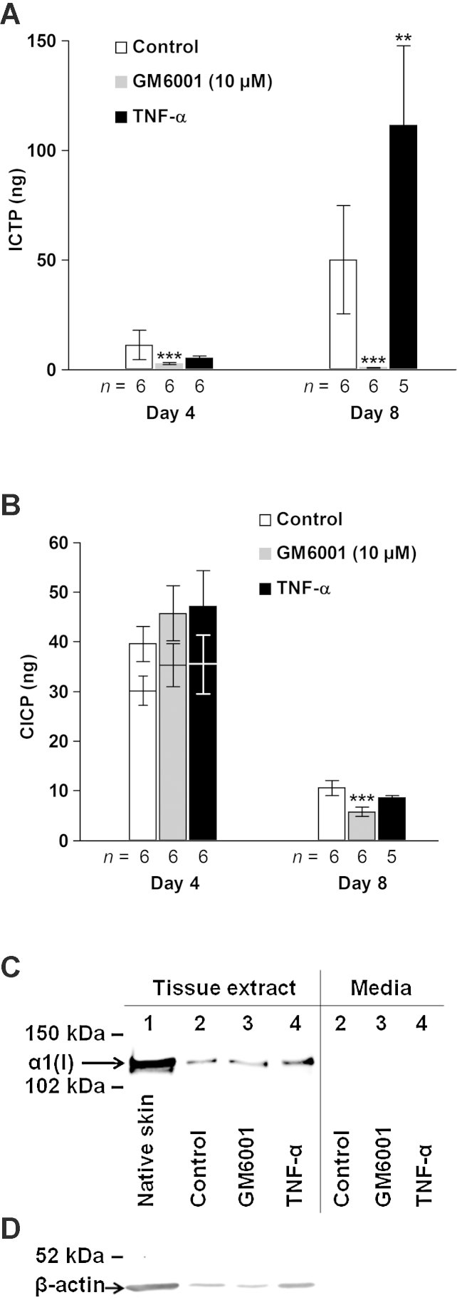
Tumor necrosis factor (TNF)-α induces matrix metalloproteinases (MMPs) that may disrupt skin integrity. We have investigated the effects and mechanisms of exogenous TNF-α on collagen degradation by incubating human skin explants in defined serum-free media with or without TNF-α (10ng/ml) in the absence or presence of the nonselective MMP inhibitor GM6001 for 8 days. The basal culture conditions promoted type I collagen catabolism that was accelerated by TNF-α (p<0.005) and accomplished by MMPs (p<0.005). Levels of the collagenases MMP-8 and MMP-13 were insignificant and neither MMP-2 nor MMP-14 were associated with increased collagen degradation. TNF-α increased secretion of MMP-1 (p<0.01) but had no impact on MMP-1 quantities in the tissue. Immunohistochemical analysis confirmed similar tissue MMP-1 expression with or without TNF-α with epidermis being the major source of MMP-1. Increased tissue-derived collagenolytic activity with TNF-α exposure was blocked by neutralizing MMP-1 monoclonal antibody and was not due to down-regulation of tissue inhibitor of metalloproteinase-1. TNF-α increased production (p<0.01), tissue levels (p<0.005) and catalytic activity of the endogenous MMP-1 activator MMP-3. Type I collagen degradation correlated with MMP-3 tissue levels (rs=0.68, p<0.05) and was attenuated with selective MMP-3 inhibitor. Type I collagen formation was down-regulated in cultured compared with native skin explants but was not reduced further by TNF-α. TNF-α had no significant effect on epidermal apoptosis. Our data indicate that TNF-α augments collagenolytic activity of MMP-1, possibly through up-regulation of MMP-3 leading to gradual loss of type I collagen in human skin.

Keywords: Aging; C-terminal telopeptide of type I collagen; Cytokine; Extracellular matrix proteins; Protease inhibitors; Type I C-terminal collagen propeptide; UK370106.


Figures

Fig. 1
Fig. 1
Effect of TNF-α on collagen degradation of cultured human skin explants monitored by hydroxyproline-containing peptides released into the media after 8 days in culture. The accumulated amount of hydroxyproline from 10 separate organ-cultured 8-mm explants of each of the 6 donors and group (control and TNF-α) was used for the global calculations. Mean ± SEM (n = 6). (For interpretation of the references to color in this figure legend, the reader is referred to the web version of this article.)
Fig. 2
Fig. 2
Effect of TNF-α and GM6001 on type I collagen turnover assessed by the biochemical markers of degradation ICTP (A), neosynthesis CICP (B) and type I collagen formation (C and D). (A and B) Media from five explants per donor were pooled and then analyzed. CICP levels after ultrafiltration of day-4 samples are indicated by lower bars (B). Mean ± SEM. **p < 0.01, ***p < 0.005 versus control at respective time point. (C and D) Western blot analysis for the α1 chain of type I collagen and β-actin of pooled concentrated (Amicon® Ultra; Millipore) CNTZ tissue extracts (C and D) and conditioned media (C) from 30 individual 8-mm skin explants (5 explants from each of the 6 donors). (C) Loading of tissue extracts was normalized to the β-actin content determined separately (D) and media were adjusted to the corresponding volume to biopsy weight ratio. Lane 1, native skin; 2, control; 3, GM6001 (10 μM); 4, TNF-α. (D) Equal volume (12.5 μl) of the tissue extracts was applied to each well.
Fig. 3
Fig. 3
LDH activity in media from cultured skin explants (A), and in cell lysates of and media from normal human epidermal keratinocytes (B). (A) Individual samples, each comprising the combined media from 5 separate 8-mm skin explants, were analyzed. n = number of skin donors. (B) Confluent keratinocytes of the second passage were treated with or without TNF-α for 2 days. Mean ± SEM. *p < 0.05, **p < 0.01, ***p < 0.005 versus control.
Fig. 4
Fig. 4
Apoptosis assessed by TUNEL immunohistofluorescence. (A) Digoxigenin-labeled 3′-OH DNA termini were detected by sheep polyclonal anti-digoxigenin antibody conjugated with fluorescein (red). Slides were mounted using medium containing 4′,6-diamidino-2-phenylindole (green). Representative sections of native (left), control-treated (middle) and TNF-α-treated (right) skin explants are shown. Epidermis is indicated by dashed line. Scale bars: 500 μm. (B) Total number of TUNEL-positive cells per epidermal area in mm2, determined in two sections from two explants from each donor by two blinded investigators by image analysis (NIS-Elements AR, Nikon), were used for the global calculations. Mean ± SEM (n = 4). (For interpretation of the references to color in this figure legend, the reader is referred to the web version of this article.)
Fig. 5
Fig. 5
Digestion of native type I collagen by tissue-derived proteinases of native human skin (A), control (A and B) and TNF-α-treated human skin (A–C), active rhMMP-1 (D–F) or trypsin (F). (A–C) Tissue extract pools from 30 individual 8-mm skin explants (5 explants from each of the 6 donors) per group were concentrated 3× (Amicon® Ultra; Millipore). S, substrate. (B–D) Enzymes were incubated for 2 h with inhibitors before the substrate was added. (D) rhMMP-1 was incubated with substrate in the absence or presence of UK370106. (A–D, F) Collagenolytic activity in percentage of type I collagen degradation is shown below each lane. (E) Effect of rhMMP-1 on collagenolysis as a function of concentration and time of incubation (inset, 1 ng/ml). (F) Trypsin (Worthington, Lakewood, NJ, USA) treatment of native or denatured (56 °C, 30 min) substrate was carried out at identical assay conditions. rhMMP-1, 2.5 ng/ml. *, position of trypsin. D, denatured.
Fig. 6
Fig. 6
MMP and TIMP contents of native skin and cultured skin explants treated without (control) or with TNF-α (10 ng/ml) were measured in pooled tissue extracts by the Quantibody® array and expressed in total amount (ng) per explant. Each pool comprised extracts made from 30 individual 8-mm skin explants (5 explants from each of the 6 donors per group). (For interpretation of the references to color in this figure legend, the reader is referred to the web version of this article.)
Fig. 7
Fig. 7
MMP-1 and MMP-3 expression in control and TNF-α-treated skin analyzed by ELISA (A and D), Western blot (B and E) and β-casein zymography (F), and MMP-1 immunohistochemistry (C) and MMP-3 correlation to ICTP (G) after 8 days of incubation. (A and D) Pooled tissue extracts and media from five explants per donor and group were assayed. Mean ± SEM. **p < 0.01, ***p < 0.005 versus control. n = number of skin donors. (B and E) The PVDF membrane was first probed with the polyclonal MMP-1 antibody (B), then stripped and reprobed with the polyclonal MMP-3 antibody (E). Lanes 1 and 3, control; 2 and 4, TNF-α; 1 and 2, pooled tissue extract (18 μl/lane) from 30 individual 8-mm skin explants from the 6 donors (1–6) per group; lanes 3 and 4, pooled media (18 μl/lane) from 24 individual 8-mm skin explants from 5 donors (media from donor 1 was lost) per group. Std.: 42.7 kDa rhMMP-1 (14 ng). Glycosylated and nonglycosylated latent and active MMP-1/MMP-3 doublets are indicated. (C) Representative sections of native (a, d and g), or cultured skin explants from control (b, e and h) and TNF-α groups (c, f and i) treated with primary monoclonal MMP-1 antibody that detects latent and active forms (a–f) or with isotype (negative) control (g–i). (a–c, 40×; d–i, 900×). (F) Casein gels were incubated in the absence (Buffer) or presence of 10 μM GM6001. Lane 1, rhMMP-3 (5 ng); 2, pooled media from control-treated explants (15 μl); 3, pooled media from TNF-α-treated explants (15 μl); 4, rhMMP-1 (2 ng). Mark12™ (Life Technologies) molecular weight marker was run in parallel lane. Upper doublets represent latent forms of MMP-3 (upper bands) and MMP-1 (lower bands) and lower doublets active forms of MMP-3 (upper bands) and MMP-1 (lower bands). (G) Tissue MMP-3 contents and corresponding ICTP in media. Each symbol as indicated in Fig. 1 represents pooled tissue extracts and media from five explants per donor. The TNF-α-treated skin explants of donor 1 is missing due to lost pooled media sample. (For interpretation of the references to color in this figure legend, the reader is referred to the web version of this article.)
Fig. 8
Fig. 8
MMP-2 (A and B) and TIMP-1 (C) tissue levels. (A) Total and active (lower bars) MMP-2 contents estimated by gelatin zymography. (B) In the zymogram, MMP-2 standard and pooled tissue extracts (1.0 μl) from 30 individual 8-mm skin explants per group were loaded into each lane. Lane 1, rhMMP-2 (50 pg; PF037); 2, native skin; 3, control; 4, TNF-α. (C) TIMP-1 levels determined by ELISA. (A and C) Pooled tissue extracts from five separate native or organ-cultured 8-mm skin explants from each of the donors and group were used for the analyses. (For interpretation of the references to color in this figure legend, the reader is referred to the web version of this article.) Mean ± SEM (n = 6).
None

Similar articles

Cited by

References

    1. Ågren M.S., Andersen T.L., Andersen L., Schiødt C.B., Surve V., Andreassen T.T. Nonselective matrix metalloproteinase but not tumor necrosis factor-alpha inhibition effectively preserves the early critical colon anastomotic integrity. Int. J. Colorectal Dis. 2011;26:329–337. - PubMed
    1. Ågren M.S., Andersen T.L., Mirastschijski U., Syk I., Schiødt C.B., Surve V. Action of matrix metalloproteinases at restricted sites in colon anastomosis repair: an immunohistochemical and biochemical study. Surgery. 2006;140:72–82. - PubMed
    1. Bashir M.M., Sharma M.R., Werth V.P. TNF-alpha production in the skin. Arch. Dermatol. Res. 2009;301:87–91. - PubMed
    1. Boström P., Söderström M., Vahlberg T., Söderström K.O., Roberts P.J., Carpén O. MMP-1 expression has an independent prognostic value in breast cancer. BMC Cancer. 2011;11:348. - PMC - PubMed
    1. Brew K., Nagase H. The tissue inhibitors of metalloproteinases (TIMPs): an ancient family with structural and functional diversity. Biochim. Biophys. Acta. 2010;1803:55–71. - PMC - PubMed

Publication types