Review
. 2014 Oct;93(10-12):367-79.
doi: 10.1016/j.ejcb.2014.10.002. Epub 2014 Oct 23.

Invadosomes in their natural habitat

Affiliations
Review

Invadosomes in their natural habitat

Elisabeth Génot et al. Eur J Cell Biol. 2014 Oct.

Abstract

Podosomes and invadopodia (collectively known as invadosomes) are small, F-actin-rich protrusions that are located at points of cell-ECM contacts and endow cells with invasive capabilities. So far, they have been identified in human or murine immune (myelomonocytic), vascular and cancer cells. The overarching reason for studying invadosomes is their connection to human disease. For example, macrophages and osteoclasts lacking Wiskott-Aldrich syndrome protein (WASp) are not able to form podosomes, and this leads to altered macrophage chemotaxis and defective bone resorption by osteoclasts. In contrast, the ability of cancer cells to form invadopodia is associated with high invasive and metastatic potentials. While invadosome composition, dynamics and signaling cascades leading to their assembly can be followed easily in in vitro assays, studying their contribution to pathophysiological processes in situ remains challenging. A number of recent papers have started to address this issue and describe invadosomes in situ in mouse models of cancer, cardiovascular disease and angiogenesis. In addition, in vivo invadosome homologs have been reported in developmental model systems such as C. elegans, zebrafish and sea squirt. Comparative analyses among different invasion mechanisms as they happen in their natural habitats, i.e., in situ, may provide an outline of the invadosome evolutionary history, and guide our understanding of the roles of the invasion process in pathophysiology versus development.

Keywords: Cancer; Cell locomotion; Cell migration; Cell motility; Confocal; In situ; In vivo; Invadopodia; Invadosomes; Invasion; Microenvironment; Microscopy; Multiphoton; Podosomes; Protrusions; development.


Figures

Figure 1
Figure 1. Endothelial cells in the murine aorta form podosome rosettes upon exposure to TGFβ ex vivo
A living aortic segment was explanted from an anesthetized mouse, opened up along its long axis, was treated with TGFβ for 20 h, then processed for fluorescence staining and analyzed by confocal microscopy for “en face” viewing. Serial optical sections are shown and presented from the apical (top) to the basal (bottom) regions of the endothelial monolayer. F-actin (red) and Hoechst 33342 (blue) distinguish between ECs with large round nuclei and vascular VSMCs with thin and elongated nuclei. The merged staining for F-actin (red) and cortactin (green) shows the rosette located at the basal surface of the ECs, extending towards the VSMC along the z-axis. Bar, 5 μm. Courtesy of Patricia Rottiers, (University of Bordeaux).
Figure 2
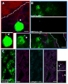
Figure 2. Presence of invadosome markers in xenografts of MDA-MB-231 human breast carcinoma
(A) A tumor cell (green) forms a small protrusion (arrow) oriented towards a blood vessel (red). Dendra2 is stably expressed in tumor cells (green); flowing blood vessels are labeled with Texas Red 70kDa dextran; collagen fibers are visualized via second harmonic generation (purple) with a multiphoton microscope. In the insert below, a small protrusion was identified using motion analysis: Images from a time lapse series taken at 30min and 0min were subtracted and the difference in the images was marked in red, revealing dynamic pixels. (B) Small protrusions in vivo contain cortactin colocalized with ECM degradation. Cortactin-GFP is stably expressed in MDA-MB-231 cells (green); ECM degradation is transiently labeled in vivo by injection of MMPSense680 (cyan); collagen fibers are shown in purple. Scale bar 20 μm. Arrow in overlay image and zoomed-in insert points to colocalization of cortactin and degradation. (C) Tks5-rich punctae colocalize with ECM degradation in situ, in tumor cryosections. Green-cytoplasmic Dendra2; purple-Tks5 mAb; cyan-degraded ECM visualized using antibody against fragments of collagen I (collagen I-¾ C, (Gligorijevic et al., 2012)). Arrows in overlay image and zoomed-in inserts point to specific points of colocalization of Tks5 and degradation. White lines delineate blood vessels. Scale bar 20 μm. Courtesy of Bojana Gligorijevic and John Condeelis (Albert Einstein College of Medicine).
Figure 3
Figure 3. Transgenic mouse model MMTV-PyMT exhibits small protrusions in perivascular regions
Tumor cells (green) in a region rich in collagen fibers (purple), macrophages (white) and blood vessels (red) in transgenic animals MMTV-PyMT × MMTV-iCre/CAG-CAC-Dendra2 at carcinoma stage (15 w). The white box is enlarged in (B), focusing on a single 5 μm-thick slice in the green channel. (B) During a 30-minute time-lapse, tumor cells next to blood vessels do not move their body but extend small protrusions (B, red overlay). All small protrusions are located at the tumor cell-blood vessel contacts (white line). Scale bars 75 μm in (A), 20 μm in (B). Protrusion extension tracked over 30 minutes is overlaid in red (B). Also see Supplementary Video 1. Courtesy of Bojana Gligorijevic and John Condeelis (Albert Einstein College of Medicine).
Figure 4
Figure 4. An F-actin-rich, invasive protrusion initiates the breach of the basement membrane during anchor cell invasion in C. elegans.
Panel is a single confocal section of the anchor cell’s surface in contact with the basement membrane (top-down-view). An F-actin probe (middle; moeABD::mCherry) specifically expressed in the anchor cell reveals a puncta located in the center of the anchor cell’s invasive cell membrane breaching the BM (right, small gap in laminin::GFP; left, overlay). Scale bar = 5μm. Courtesy of Elliott J. Hagedorn and David R. Sherwood (Duke University).
Figure 5
Figure 5. Zebrafish embryonic development of neural crest and intestine requires Tks5-dependent protrusions
(A) Neural crest cells labeled with transgenic (sox10:RFP) in control (T5 MM) and Tks5 morphant (T5 MO) (30 hours post-fertilization, hpf). Arrows mark individual cells which migrate over the 1.5 hours duration. * = protrusions emanating from neural crest cells. Image reproduced from Murphy et al, 2011. (B) Actin-rich protrusions in epithelial cells of the intestine in meltdown mutant (mlt). 3D rendering of sagittal confocal sections through the intestine of 78 hpf wild type (WT) and mlt larvae. Actin is labeled by transgenic Lifeact-GFP expression (green). In WT, actin is mainly in the epithelial cell apical brush border (bracket). In mlt, actin-rich protrusions are detected (arrows), in addition to brush border actin (bracket). (C) Actin-rich protrusions in mlt co-localize with sites missing basal lamina (lower panel, arrowhead and inset). Histological cross-sections through the intestine of 74 hpf immunostained mlt larvae. BM is labeled red (laminin Ab), actin is green (GFP Ab) and nuclei are blue (DAPI). ap, apical epithelial cell borders; ba, basal cell epithelial cell border. Images (B, C) reproduced from Seiler et al., 2012.

References

    1. Albinsson S, Sward K. Targeting smooth muscle microRNAs for therapeutic benefit in vascular disease. Pharmacol Res. 2013;75:28–36. - PubMed
    1. Alexander NR, Branch KM, Parekh A, Clark ES, Iwueke IC, Guelcher SA, Weaver AM. Extracellular Matrix Rigidity Promotes Invadopodia Activity. Curr Bio. 2009;18:1295–1299. - PMC - PubMed
    1. Annes JP, Munger JS, Rifkin DB. Making sense of latent TGFβ activation. J Cell Sci. 2003;116:217–24. - PubMed
    1. Artym VV, Zhang Y, Seillier-Moiseiwitsch F, Yamada KM, Mueller SC. Dynamic interactions of cortactin and membrane type 1 matrix metalloproteinase at invadopodia: defining the stages of invadopodia formation and function. Cancer Res. 2006;66:3034–43. - PubMed
    1. Ayala I, Giacchetti G, Caldieri G, Attanasio F, Mariggiò S, Tetè S, Polishchuk R, Castronovo V, Buccione R. Faciogenital dysplasia protein Fgd1 regulates invadopodia biogenesis and extracellular matrix degradation and is up-regulated in prostate and breast cancer. Cancer Res. 69:747–52. - PubMed

Publication types

MeSH terms

LinkOut - more resources