. 2014 Nov 24;31(4):393-404.
doi: 10.1016/j.devcel.2014.10.014. Epub 2014 Nov 24.

RanBP1 governs spindle assembly by defining mitotic Ran-GTP production

Affiliations

RanBP1 governs spindle assembly by defining mitotic Ran-GTP production

Michael Shaofei Zhang et al. Dev Cell. .

Abstract

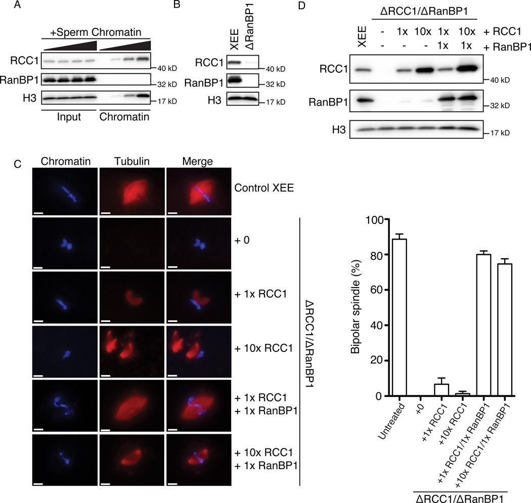
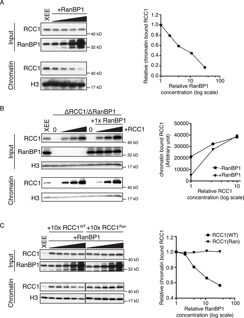
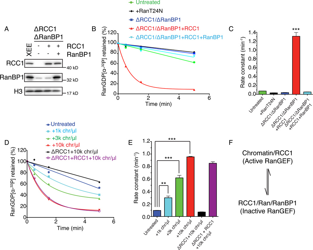
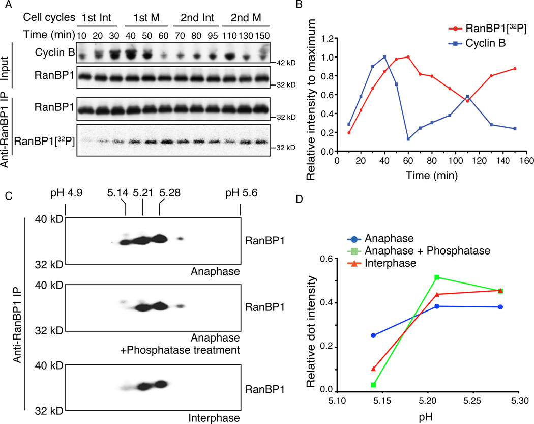
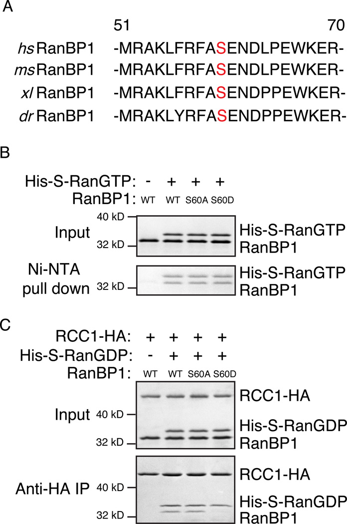
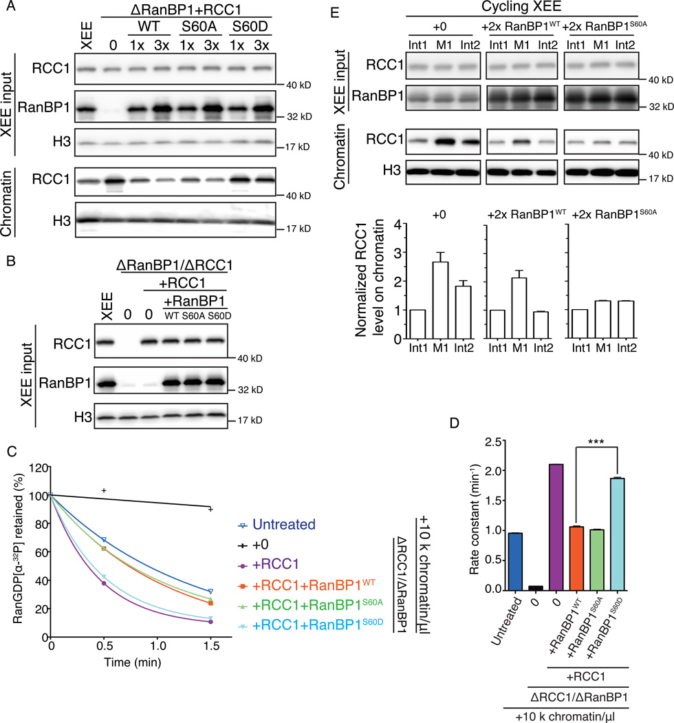
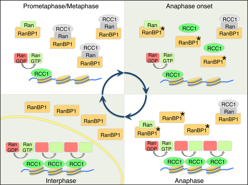
Accurate control of the Ras-related nuclear protein (Ran) GTPase cycle depends on the regulated activity of regulator of chromosome condensation 1 (RCC1), Ran's nucleotide exchange factor. RanBP1 has been characterized as a coactivator of the Ran GTPase-activating protein RanGAP1. RanBP1 can also form a stable complex with Ran and RCC1, although the dynamics and function of this complex remain poorly understood. Here, we show that formation of the heterotrimeric RCC1/Ran/RanBP1 complex in M phase Xenopus egg extracts controls both RCC1's enzymatic activity and partitioning between the chromatin-bound and soluble pools of RCC1. This mechanism is critical for spatial control of Ran-guanosine triphosphate (GTP) gradients that guide mitotic spindle assembly. Moreover, phosphorylation of RanBP1 drives changes in the dynamics of chromatin-bound RCC1 pools at the metaphase-anaphase transition. Our findings reveal an important mitotic role for RanBP1, controlling the spatial distribution and magnitude of mitotic Ran-GTP production and thereby ensuring accurate execution of Ran-dependent mitotic events.


Figures

Figure 1
Figure 1. RanBP1 is required in normal spindle assembly by sequestering RCC1
(A) Different concentrations (0, 1,000, 3,000 and 10,000 units/µl) of demembraned sperm chromatin were added and re-purified from each sample. Total CSF-XEE input (left half) and isolated chromatin (right half) were subjected to immunblottinged with antibodies against RCC1, RanBP1 and Histone H3. (B) Mock treated CSF-XEE (left) or CSF-XEE depleted with anti-RanBP1 antibodies (right) were examined by immunoblotting with antibodies against RCC1, RanBP1 and Histone H3. (C) Recombinant xRCC1 and/or xRanBP1 were added in to mock treated CSF-XEE or CSF-XEE depleted using anti-RanBP1 antibodies. Rhodamine-labeled α-tubulin (20 µg/ml) and demembraned chromatin (1,000 units/µl) were added. After 30 minutes at RT, aliquots of each reaction were fixed, stained with Hoechst 33342 and processed for fluorescent microscopy. Images were taken for chromatin (left, blue) and tubulin (middle, red). Scale bar, 10 µm. Spindles with chromatin in vicinity were counted as one structure. Percentage of bipolar spindles with chromatin correctly localizing at the mid-plate was plotted as mean ± SEM (N = 3 XEEs, 50 structures counted in each XEE). (D) Samples as in (C) were subjected to immunoblotting with antibodies against RCC1, RanBP1, and Histone H3. See also Figure S1.
Figure 2
Figure 2. RanBP1 competes with chromatin in binding RCC1
(A) Increasing concentrations of recombinant xRanBP1 (0×, 1×, 3×, 10×, 30× endogenous RanBP1 level) were added to CSF-XEE. Reaction aliquots (upper panels) and isolated chromatin (lower panels) were subjected to immunoblotting with indicated antibodies. Relative chromatin bound RCC1 was plotted against RanBP1 concentration (folds of endogenous level). (B) Recombinant xRCC1 was added at increasing concentrations (0×, 1×, 3×, 10× relative to endogenous RCC1) to CSF-XEE previously depleted using anti-RanBP1 antibodies. Each reaction was divided, and either buffer (left) or recombinant RanBP1 (right) were added. Reaction aliquots (upper panels) and isolated chromatin (lower panels) were subjected to immunoblotting with indicated antibodies. Chromatin bound RCC1 was plotted against RCC1 concentration (folds of endogenous level). (C) Either recombinant wild type RCC1 (RCC1WT) or an RCC1 mutant that does not bind Ran (RCC1Ran) was added into CSF-XEE at 10× endogenous RCC1 levels. Recombinant xRanBP1 was added to either sample at increasing concentrations (0×, 1×, 3×, 10×, 30× relative to endogenous RanBP1). Reaction aliquots (upper panels) and isolated chromatin (lower panels) were subjected to immunoblotting with indicated antibodies. Relative chromatin bound RCC1 was plotted against RanBP1 concentration (folds of endogenous level). In all panels, all reactions contained 10,000 units/µl demembraned sperm chromatin, and were incubated for 30 min at RT before being re-purified. See also Figure S2.
Figure 3
Figure 3. RanBP1 complex inhibited RCC1 in mitotic cytosol
(A) RCC1 and RanBP1 were depleted from CSF-XEE using anti-RanBP1 antibodies (lane 2–4). Buffer (lane 2) or physiological concentrations of recombinant xRCC1 (lane 3) or of recombinant xRCC1 and xRanBP1 (lane 4) were added back to the depleted CSF-XEE. Total CSF-XEE was subjected to immunblotting by indicated antibodies. Lane 1 shows undepleted control CSF-XEE. (B) 10 µM recombinant Ran charged with [α-32P]GDP was added to CSF-XEE reactions as in (A), or to undepleted CSF-XEE containing 10 µM Ran-T24N. Aliquots were taken at 0.5, 1.5, and 5 minutes after [α-32P]Ran-GDP addition, and exchange was monitored using a filter binding assay. (C) Rate constants were determined from (B) and plotted as mean ± SEM. Significance in difference was tested by two-tailed t-test (n=3, *** p<0.0001). (D) Demembraned sperm chromatin was added at the indicated concentrations to CSF-XEE, CSF-XEE depleted with anti-RCC1 antibodies, or RCC1-depleted CSF-XEE with recombinant xRCC1 (1× physiological concentration). Aliquots were sampled and analyzed as in (B). (E) Rate constants were determined from (D) and plotted as mean ± SEM. Significance in difference was tested by two-tailed t-test (n=3, ** p<0.002, *** p<0.0005). (F) RCC1 distribution equilibrium. Chromatin-bound RCC1 is an active RanGEF, while RCC1 within the RRR complex in mitotic cytosol is inactive. These two pools are in a dynamic equilibrium, determined by RanBP1 concentrations.
Figure 4
Figure 4. RanBP1 is phosphorylated on cell cycle basis
(A) 100 µCi/ml [γ-32P]ATP was added to cycling XEE. RanBP1 was immunoprecipitated from the reaction at different times and subjected to SDS-PAGE. XEE aliquots (upper panels) and RanBP1 immunoprecipitates (lower panel, row 1) were subjected to immunoblotting with indicated antibodies and 32P within the RanBP1 immunoprecipitate was detected by autoradiography (lower panel, row 2). (B) The immunoblot intensity of Cyclin B for each sample from (A) was measured and normalized relative to the maximum Cyclin B intensity (blue line). 32P associated with immunoprecipitated RanBP1 for each sample was similarly measured and normalized values for each time point are shown (red line). (C) RanBP1 was immunoprecipitated from cycling XEE at anaphase onset or interphase (bottom panel). RanBP1 precipitated from anaphase was treated with either buffer only (top panel) or alkaline phosphatase (middle panel). The RanBP1 within each sample was separated by linear pH 4–7 isoelectric focusing (IEF) followed by SDS-PAGE, and subjected to immunoblotting with antibodies against RanBP1. The numbers above show the estimated isoelectric point (pI) of individual dot. (D) Relative RanBP1 intensity for each major spot in (C) was calculated by dividing intensity of each dot to total RanBP1 intensity. The relative RanBP1 intensity of anti-RanBP1 IP from anaphase (blue line), anti-RanBP1 IP from anaphase followed by CIP treatment (green line), and anti-RanBP1 IP from interphase (red line) were plotted against pI of each dot from (C). See also Figure S3.
Figure 5
Figure 5. RanBP1 Ser 60 phosphorylation releases RCC1 to bind chromatin
(A) Protein sequence alignment of RanBP1 from H.sapiens, M.musculus, X.laevis, and D.rerio. The numbers indicate the respective positions of the first and last amino acid residue within H. sapiens RanBP1. Serine 60 is in red. (B) Recombinant His6-S-Ran-GTP was incubated with recombinant xRanBP1WT, xRanBP1S60A, or xRanBP1S60D. Complexes formed by His6-S-Ran-GTP were precipitated using Ni-NTA resin. The original reactions (upper panel) and precipitated proteins (lower panel) were separated by SDS-PAGE and visualized by Commassie Brilliant Blue (CBB) staining. As a control, a reaction was incubated in parallel, containing recombinant xRanBP1WT but lacking His6-S-Ran-GTP (lane 1). (C) Recombinant xRCC1-HA and His6-S-Ran-GDP were allowed to bind, followed by the addition of recombinant xRanBP1WT, xRanBP1S60A or xRanBP1S60D and further incubation. xRCC1-HA and associated proteins were precipitated using anti-HA beads. The original reactions (upper panel) and precipitated proteins (lower panel) were separated by SDS-PAGE and visualized by CBB staining. As a control, a reaction was incubated in parallel, containing xRCC1-HA and xRanBP1WT but lacking His6-S-Ran-GDP (lane 1). See also Figure S4.
Figure 6
Figure 6. Endogenous RanGEF activity in CSF-XEE is increased by RanBP1 phosphorylation on Ser 60
(A) xRCC1 was restored to physiological levels in CSF-XEE depleted using anti-RanBP1 antibodies by adding recombinant xRCC1. Recombinant xRanBP1WT, xRanBP1S60A or xRanBP1S60D were added as indicated (1× or 3× endogenous RanBP1 levels). 10,000 units/µl demembraned sperm chromatin were added to each sample and incubated for 30 min at RT before chromatin isolation. Each total reaction (upper panel) and the isolated chromatin (lower panel) was subjected to immunblotting with antibodies against RCC1, RanBP1 and Histone H3, as indicated. (B) CSF-XEE was depleted using anti-RanBP1 antibodies, followed by addition of buffer or physiological levels of xRCC1. Where indicated, recombinant xRanBP1WT, xRanBP1S60A or xRanBP1S60D were added at concentrations equivalent to endogenous RanBP1. Each sample was subjected to immunblotting with antibodies against RCC1, RanBP1 and Histone H3. (C) 10 µM recombinant Ran charged with [α-32P]GDP was added CSF-XEE reactions as in (B) containing demembraned sperm chromatin (10,000 units/µl). Aliquots were taken at 0.5, 1.5, and 5 minutes after [α-32P]Ran-GDP addition, and exchange was monitored using a filter binding assay, and plotted as described in Figure 3. (D) Rate constants were determined from nucleotide release data (C) and plotted as mean ± SEM. Significance in difference was tested by two-tailed t-test (n=3, *** p<0.0001). (E) Cycling XEE containing demembraned sperm chromatin (3,000 units/µl) was warmed to RT and allowed to initiate NE assembly. After NE closure, buffer (left), recombinant xRanBP1WT (middle) or xRanBP1S60A (right) were added at roughly twice the endogenous RanBP1 level. Chromatin was prepared from each sample at the first interphase (Int1), anaphase onset of the first mitosis (M1), and the second interphase (Int2). Total cycling XEE input at these three points was subjected to immunblotting with antibodies against RCC1 and RanBP1 (Row 1 and 2). Isolated chromatin from each sample at these three points was subjected to immunblotting by antibodies against RCC1 and Histone H3 (Row 3 and 4). Chromatin-bound RCC1 levels were normalized to the RCC1 signal intensity at Int1 for individual sample. The normalized RCC1 level on chromatin was plotted as mean ± SEM (n=3) (Lower panel).
Figure 7
Figure 7. Model for mitotic regulation of RCC1 by RanBP1
Premetaphase: RCC1 is partitioned between an active, chromatin bound pool (green) and an inactive pool (grey), associated with RRR complexes that also contain RanBP1 and nucleotide-free Ran (grey). Anaphase Onset: Phosphorylation of RanBP1 on Ser 60 (asterisk) releases RCC1 from the RRR complex. The free RCC1 is then recruited to chromatin. Anaphase: The high level of chromatin-bound RCC1 promotes enhanced levels of Ran-GDP (red) to Ran-GTP (green) exchange on chromatin. Interphase: NE (yellow) formation physically separates RCC1 from RanBP1, preventing RRR complex assembly and inhibition of RCC1.

Similar articles

Cited by

References

    1. Amato R, Scumaci D, D'Antona L, Iuliano R, Menniti M, Di Sanzo M, Faniello MC, Colao E, Malatesta P, Zingone A, et al. Sgk1 enhances RANBP1 transcript levels and decreases taxol sensitivity in RKO colon carcinoma cells. Oncogene. 2013;32:4572–4578. - PubMed
    1. Arnaoutov A, Dasso M. The Ran GTPase regulates kinetochore function. Developmental cell. 2003;5:99–111. - PubMed
    1. Beddow AL, Richards SA, Orem NR, Macara IG. The Ran/TC4 GTPase-binding domain: identification by expression cloning and characterization of a conserved sequence motif. Proceedings of the National Academy of Sciences of the United States of America. 1995;92:3328–3332. - PMC - PubMed
    1. Bischoff FR, Gorlich D. RanBP1 is crucial for the release of RanGTP from importin beta-related nuclear transport factors. FEBS letters. 1997;419:249–254. - PubMed
    1. Bischoff FR, Krebber H, Smirnova E, Dong W, Ponstingl H. Co-activation of RanGTPase and inhibition of GTP dissociation by Ran-GTP binding protein RanBP1. The EMBO journal. 1995;14:705–715. - PMC - PubMed

Publication types

MeSH terms

LinkOut - more resources