. 2014 Dec 9;3(6):1118-31.
doi: 10.1016/j.stemcr.2014.10.008. Epub 2014 Nov 13.

Fast and efficient neural conversion of human hematopoietic cells

Affiliations

Fast and efficient neural conversion of human hematopoietic cells

Julio Castaño et al. Stem Cell Reports. .

Abstract

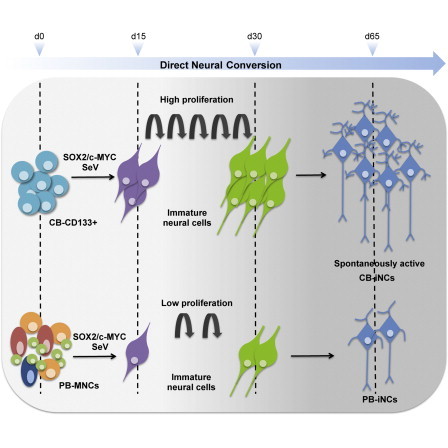
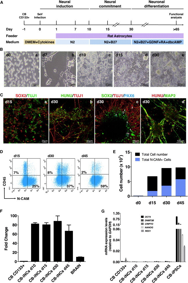
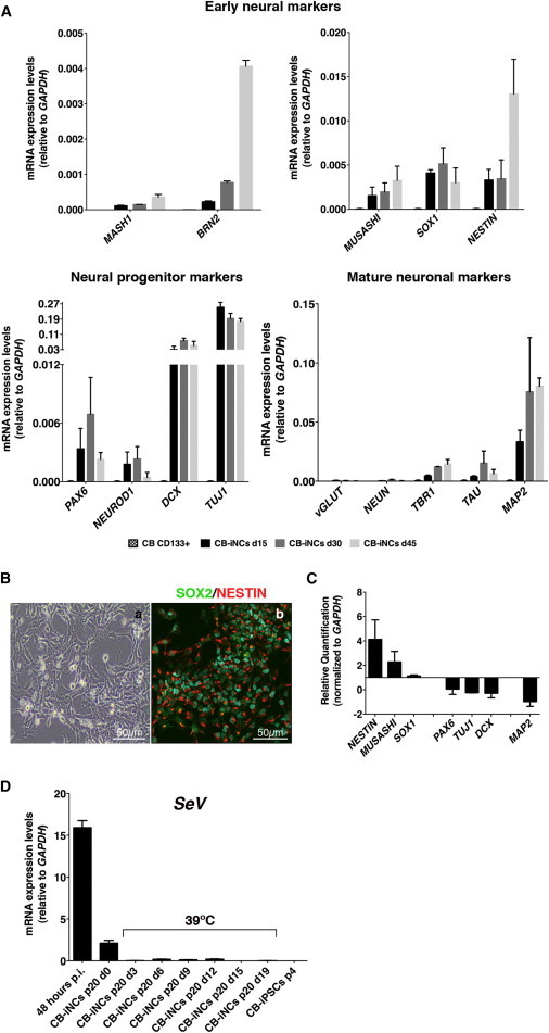
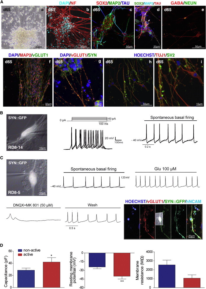
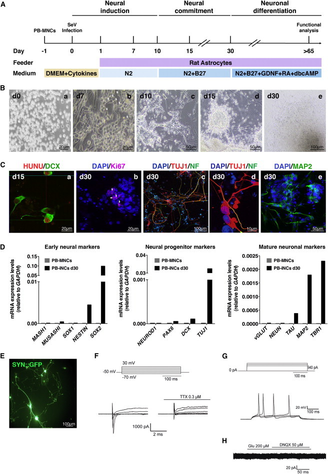
Neurons obtained directly from human somatic cells hold great promise for disease modeling and drug screening. Available protocols rely on overexpression of transcription factors using integrative vectors and are often slow, complex, and inefficient. We report a fast and efficient approach for generating induced neural cells (iNCs) directly from human hematopoietic cells using Sendai virus. Upon SOX2 and c-MYC expression, CD133-positive cord blood cells rapidly adopt a neuroepithelial morphology and exhibit high expansion capacity. Under defined neurogenic culture conditions, they express mature neuronal markers and fire spontaneous action potentials that can be modulated with neurotransmitters. SOX2 and c-MYC are also sufficient to convert peripheral blood mononuclear cells into iNCs. However, the conversion process is less efficient and resulting iNCs have limited expansion capacity and electrophysiological activity upon differentiation. Our study demonstrates rapid and efficient generation of iNCs from hematopoietic cells while underscoring the impact of target cells on conversion efficiency.


Figures

None
Graphical abstract
Figure 1
Figure 1
Generation of iNCs from CD133-Positive CB Cells Using SeV Vectors (A) Schematic timeline of neural conversion. CD133-positive CB cells transduced with SOX2 and c-MYC SeV vectors were seeded on irradiated rat astrocytes and cultured with N2 medium supplemented with LDN, CHIR99021, and SB431542 compounds for 10 days. From days 10 to 30, cells were cultured in N2 + B27 medium. From day 30 onward, CB-iNCs were terminally differentiated in the presence of N2 + B27 medium with BDNF, GDNF, RA, and dbcAMP. Electrophysiology was performed at day 65. (B) Representative phase-contrast images illustrating the morphological conversion of CD133-positive CB cells into iNCs. Note that cells with neural progenitor morphology were identifiable already at day 7 (n = 5 independent experiments). Scale bars are as indicated in the panels. (C) Representative immunofluorescence images of CB-iNCs at days 15 and 30. Two weeks after induction, CB-iNCs were SOX2 positive and TUJ1 positive and exhibited a neural morphology (a). By day 30, CB-iNCs showed a more complex cytoarchitecture, organized in clusters with long TUJ1- and MAP2-positive processes (b–d). Of note, few PAX6-positive cells (white arrows) were observed (c). Scale bars are as indicated in the panels. (D) Flow cytometry quantification of CD45 and CD56 (N-CAM) in a representative experiment, showing the progressive increase in N-CAM-positive cells reaching ∼60% by day 45. (E) Quantification of total cell number and N-CAM-positive neuronal cells at differentiation days 15, 30, and 45. (F) Quantification of endogenous SOX2 mRNA levels in CB-iNCs by qRT-PCR analysis. Data are shown as fold induction of SOX2 mRNA level in CB-iNCs relative to CD133-positive CB cells, which is considered as 1. Human total brain RNA (YORBIO) was used as positive control. Data are represented as mean ± SD (n = 3 independent experiments). (G) Pluripotency genes (OCT4, NANOG, REX1, CRIPTO, and DNMT3B) expression was not detected in CB-iNCs by qRT-PCR during the conversion process. CB derived iPSCs were used as positive control. Data are represented as mean ± SD (n = 3 independent experiments). See also Figure S1.
Figure 2
Figure 2
Characterization iNCs Derived from CD133-Positive CB Cells (A) Quantification of expression levels by qRT-PCR of a panel of early, immature, and mature neuronal markers in CD133-positive CB cells and CB-iNCs at 15, 30, and 45 days of neural induction and propagation. Data are represented as mean ± SD (n = 3 independent experiments). Expression of these genes was not detected in untransduced CD133-positive CB cells. (B) Representative phase-contrast live image (a) and immunostaining (b) of CB-iNCs at passage 5 cultured on matrigel in the presence of FGF2. CB-iNCs acquired a neural epithelial-like morphology and expressed early neural progenitor markers such as NESTIN and SOX2. The scale bar is as indicated in the panels. (C) qRT-PCR analysis showing transcriptional changes in the CB-iNCs propagated on matrigel in the presence of FGF2 for five passages relative to CB-iNCs at day 30 of neural induction. Data are represented as mean ± SD (n = 3 independent experiments). (D) SeV vectors analyzed by qRT-PCR after 20 passages before and after temperature treatment (39°C)-mediated silencing. Data are represented as mean ± SD (n = 3 independent experiments). See also Figure S2.
Figure 3
Figure 3
In Vitro Neuronal Differentiation and Whole-Cell Patch-Clamp Recordings of CB-iNCs (A) Representative phase contrast (a) and immunofluorescence (b–i) images of CB-iNCs at differentiation day 65. Differentiated CB-iNCs expressed postmitotic mature markers such as axonal specific neurofilament SMI 312 (NF), MAP2, TAU (T46 epitope), and NEUN (RBFOX3) (b–e). (d) High-magnification image of TAU-positive cells showing downregulation of SOX2 nuclear staining, indicated by white arrows. CB-iNCs can differentiate into inhibitory GABA-positive neurons (e) and excitatory vGLUT1-positive neurons (f). Mature neurons derived from CB-iNCs after 65 days of differentiation expressed synaptic proteins, such as SYN and SV2 (g–i). Scale bars are as indicated in the panels. (B) Current-clamp recordings from a GFP-positive (SYN::GFP) cell showing evoked action potentials by injecting 20 pA current steps (middle panel) and spontaneous action potentials at a −40 mV resting membrane potential (I = 0) (right panel). The scale bar is as indicated in the panel. (C) Current-clamp recordings of another GFP-positive (SYN::GFP) cell in the same culture showing spontaneous activity that was pharmacologically modulated by glutamate (upper right panel) and glutamate antagonists (DNQX: non-NMDA receptor antagonist, MK801: NMDA receptor antagonist, left and middle bottom panels). Representative traces of spontaneous activity are truncated for clarity. Immunofluorescence confirmed expression of vGLUT1, together with GFP (SYN::GFP) and human specific N-CAM in this cell (right bottom panel). Scale bars are as indicated in the panels. (D) Spontaneously active cells showed more mature electrophysiological properties, as capacitance was significantly higher (t35 = 1.66, p = 0.05) (left panel) and the resting membrane potential significantly lower (t29 = 2.51, p < 0.01) (middle panel) than those recorded from nonactive cells. Membrane resistance was smaller, although not statistically different (t35 = 1.45, p = 0.07) (right panel). p ≤ 0.05; ∗∗p < 0.01; unpaired t test. See also Figure S3.
Figure 4
Figure 4
Generation and Characterization of iNCs from PB-MNCs Using SeV (A) Schematic representation of neural induction of PB-MNCs. Two hundred fifty thousand PB-MNCs were transduced with SOX2 and c-MYC SeV vectors at high MOI. At day 1, infected cells were plated on irradiated rat astrocytes in the presence of N2 medium supplemented with LDN, CHIR99021, and SB431542 compounds. From days 10 to 30, cells were cultured in N2 + B27 medium. From day 30 onward, PB-iNCs were terminally differentiated in the presence of N2 + B27 medium with BDNF, GDNF, RA, and dbcAMP for another 6–8 weeks. (B) Representative phase contrast images illustrating the time course of the conversion of PB-MNCs into neural cells. Similar to CB, some neuroepithelial cells (indicated by white arrows) emerged as early as day 7 (n = 4 independent experiments). Scale bars are as indicated in the panels. (C) Representative immunofluorescence images of converted PB-iNCs at days 15 and 30. At day 15, PB-iNCs were DCX positive and showed an immature morphology; (a) is a confocal orthogonal reconstruction to ensure colocalization with human nuclear antigen HUNU. There were few Ki67-positive cells (indicated by white arrows) in concordance with the limited expansion capacity (b). By day 30, PB-iNCs displayed a more mature morphology and expressed TUJ1, axonal specific neurofilament (NF), and MAP2 (c–e). Scale bars are as indicated in the panels. (D) Representative qRT-PCR analysis of early, immature, and mature neuronal markers in PB-MNCs and PB-iNCs after 30 days of neural induction. Note that expression of these genes was not detected in untransduced PB-MNCs. (E) PB-iNCs in culture expressing SYN::GFP after lentiviral transduction. GFP-positive cells were patched for electrophysiology. The scale bar is as indicated in the panel. (F) Inward Na+ and outward K+ voltage-dependent currents triggered upon −70 mV to +50 mV voltage steps (Vh = −50 mV). Na+ currents were TTX sensitive. Representative traces of voltage-dependent currents were truncated and expanded for clarity. (G) PB-iNCs GFP-positive cells showed action potentials evoked by somatic current injection (step 20 pA, n = 15). (H) No current changes were observed in response to either glutamate or the glutamate receptor antagonist DNQX (Vh = −50 mV). See also Figure S4.

References

    1. Ambasudhan R., Talantova M., Coleman R., Yuan X., Zhu S., Lipton S.A., Ding S. Direct reprogramming of adult human fibroblasts to functional neurons under defined conditions. Cell Stem Cell. 2011;9:113–118. - PMC - PubMed
    1. Arnold K., Sarkar A., Yram M.A., Polo J.M., Bronson R., Sengupta S., Seandel M., Geijsen N., Hochedlinger K. Sox2(+) adult stem and progenitor cells are important for tissue regeneration and survival of mice. Cell Stem Cell. 2011;9:317–329. - PMC - PubMed
    1. Avilion A.A., Nicolis S.K., Pevny L.H., Perez L., Vivian N., Lovell-Badge R. Multipotent cell lineages in early mouse development depend on SOX2 function. Genes Dev. 2003;17:126–140. - PMC - PubMed
    1. Ban H., Nishishita N., Fusaki N., Tabata T., Saeki K., Shikamura M., Takada N., Inoue M., Hasegawa M., Kawamata S., Nishikawa S. Efficient generation of transgene-free human induced pluripotent stem cells (iPSCs) by temperature-sensitive Sendai virus vectors. Proc. Natl. Acad. Sci. USA. 2011;108:14234–14239. - PMC - PubMed
    1. Bellin M., Marchetto M.C., Gage F.H., Mummery C.L. Induced pluripotent stem cells: the new patient? Nat. Rev. Mol. Cell Biol. 2012;13:713–726. - PubMed

Publication types