. 2014 Nov 19;84(4):681-96.
doi: 10.1016/j.neuron.2014.09.007. Epub 2014 Nov 19.

Studying brain organization via spontaneous fMRI signal

Affiliations

Studying brain organization via spontaneous fMRI signal

Jonathan D Power et al. Neuron. .

Abstract

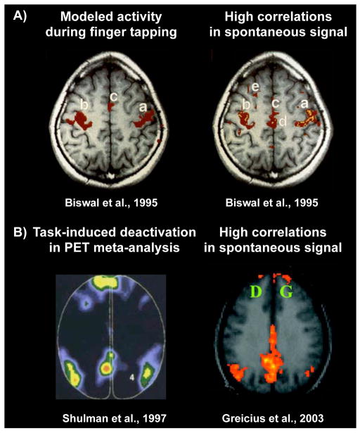
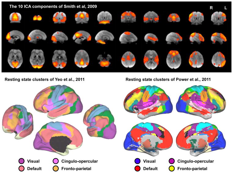
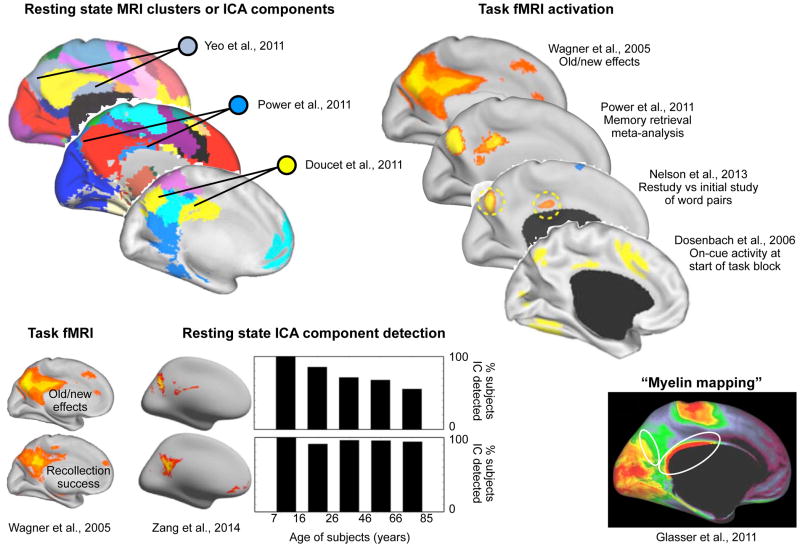
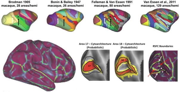
In recent years, some substantial advances in understanding human (and nonhuman) brain organization have emerged from a relatively unusual approach: the observation of spontaneous activity, and correlated patterns in spontaneous activity, in the "resting" brain. Most commonly, spontaneous neural activity is measured indirectly via fMRI signal in subjects who are lying quietly in the scanner, the so-called "resting state." This Primer introduces the fMRI-based study of spontaneous brain activity, some of the methodological issues active in the field, and some ways in which resting-state fMRI has been used to delineate aspects of area-level and supra-areal brain organization.


Conflict of interest statement

The authors have no conflict of interest to report.

Figures

Figure 1
Figure 1. Spatial correspondence between task-evoked activity patterns and patterns in spontaneous fMRI signal correlations
A) Modeled BOLD activity during finger tapping, and a seed correlation map with location (a) as the seed. B) Locations in a PET meta-analysis where deactivations are seen across tasks, and a seed correlation map with the posterior cingulate as the seed. Images modified from the indicated publications.
Figure 2
Figure 2. Large-scale correlation patterns in resting state fMRI data
Data are shown from 3 reports on the spatial patterns of correlated BOLD signal: a 10-component ICA analysis, a surface-based analysis of surface vertex clustering, and a volume-based analysis of voxelwise clustering. Images modified from the indicated publications.
Figure 3
Figure 3. A medial parietal resting state network exhibits specific functional, structural, and lifespan properties
A grouping of regions mainly in medial parietal cortex that has been identified in multiple resting state analyses exhibits memory-related and oldness-related activations, activity at the beginning of a task block, specific age-related decreases in resting state correlations, and increased putative myelin content relative to surrounding tissue. Images modified from the indicated publications. Note that the illustration from (Yeo et al., 2011) is the 17-cluster partitioning, not the 7-cluster partitioning shown in Figure 2.
Figure 4
Figure 4. Area-level mapping of cortex in macaques and humans
At top, several macaque cortical parcellation schemes show the refinement of area-level maps over the last century, modified from (Van Essen et al., 2012a). At bottom, a parcellation of the human cortex based on resting state functional connectivity. The insets show postero-medial views of the occipital lobe of the left hemisphere, with cytoarchitectonic locations for Brodmann areas 17 and 18, and corresponding resting state boundaries. The white arrows denote borders that align well with the predicted area borders, and the red arrows denote boundaries that may be byproducts of unremoved artifact at the occipital pole. Modified from (Wig et al., 2014b).
Figure 5
Figure 5. Considerations in resting state networks: node definition
The 90-parcel AAL atlas is shown, as are seed maps from sites within two of the parcels. The clustering structure of resting state networks with nodes of voxels, functionally-defined ROIs, and the AAL parcels are shown. Image at left is modified from www.prefrontal.org and the images at right are modified from (Power et al., 2011).

References

    1. Adachi Y, Osada T, Sporns O, Watanabe T, Matsui T, Miyamoto K, Miyashita Y. Functional connectivity between anatomically unconnected areas is shaped by collective network-level effects in the macaque cortex. Cereb Cortex. 2012;22:1586–1592. - PubMed
    1. Amunts K, Lepage C, Borgeat L, Mohlberg H, Dickscheid T, Rousseau ME, Bludau S, Bazin PL, Lewis LB, Oros-Peusquens AM, et al. BigBrain: an ultrahigh-resolution 3D human brain model. Science. 2013;340:1472–1475. - PubMed
    1. Anderson JS, Druzgal TJ, Lopez-Larson M, Jeong EK, Desai K, Yurgelun-Todd D. Network anticorrelations, global regression, and phase-shifted soft tissue correction. Hum Brain Mapp. 2011;32:919–934. - PMC - PubMed
    1. Barnes KA, Nelson SM, Cohen AL, Power JD, Coalson RS, Miezin FM, Vogel AC, Dubis JW, Church JA, Petersen SE, Schlaggar BL. Parcellation in left lateral parietal cortex is similar in adults and children. Cereb Cortex. 2012;22:1148–1158. - PMC - PubMed
    1. Beauchamp MS, Argall BD, Bodurka J, Duyn JH, Martin A. Unraveling multisensory integration: patchy organization within human STS multisensory cortex. Nat Neurosci. 2004;7:1190–1192. - PubMed

Publication types

LinkOut - more resources