Review
. 2015 Feb;35(1):161-83.
doi: 10.1016/j.iac.2014.09.008. Epub 2014 Nov 21.

Atopic dermatitis in children: clinical features, pathophysiology, and treatment

Affiliations
Review

Atopic dermatitis in children: clinical features, pathophysiology, and treatment

Jonathan J Lyons et al. Immunol Allergy Clin North Am. 2015 Feb.

Abstract

Atopic dermatitis (AD) is a chronic, relapsing, highly pruritic skin condition resulting from disruption of the epithelial barrier and associated immune dysregulation in the skin of genetically predisposed hosts. AD generally develops in early childhood, has a characteristic age-dependent distribution and is commonly associated with elevated IgE, peripheral eosinophilia, and other allergic diseases. Medications such as antihistamines have demonstrated poor efficacy in controlling AD-associated itch. Education of patients regarding the primary underlying defects and provision of a comprehensive skin care plan is essential for disease maintenance and management of flares.

Keywords: Allergy; Atopic dermatitis; Eczema; Hyper-IgE syndrome; Netherton syndrome.


Figures

Fig 1
Fig 1
Typical distribution of skin lesions in a child with atopic dermatitis.
Fig 2
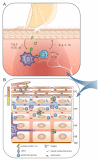
Fig 2. Defective pathways contributing to the pathogenesis of atopic dermatitis
(A) Intense pruritus develops in lesional skin; defective barrier function leads to immune activation by irritants and microbes, with IL-25, IL-33, and TSLP contributing to Th2-mediated inflammation. IL-4 and IL-13 induce class switching to IgE, and further impair skin barrier function by reducing both filaggrin and anti-microbial peptide expression. Highly pruritogenic IL-31 is also elaborated leading to additional excoriation, further skin barrier degradation and immune activation, thus potentiating disease. (B) Mutations in several proteins expressed in the epidermis result in severe atopic dermatitis; these include filaggrin (FLG), lympho-epithelial Kazal-type-related inhibitor (LEKTI), as well as corneodesmosin and desmoglein-1 (components of desmosomes). FLG is critical to skin barrier integrity and hydration. Inactive profilaggrin polymers exist inside keratohyalin granules within the stratum granulosum (SG) layer. As epithelial cells transition into the stratum corneum (SC), adjacent proteases (eg. KLK5) are released by LEKTI in a pH dependent manner, acting on these granules to yield FLG monomers which contribute to skin barrier function, as well as on desmosomes to facilitate normal exfoliation. Loss of LEKTI function results in inappropriate protease activity deep within the SG resulting in the severe skin barrier defect characteristic of Netherton syndrome. Similar disease results from absence of proteins necessary for desmosome function. Further proteolysis of FLG releases small hygroscopic molecules and free amino acids that comprise Natural Moisturizing Factor (NMF). Decreased FLG results from null mutations or Th2 mediated inflammation, and leads to impaired skin hydration, dysregulated pH, and barrier dysfunction. SS – stratum spinosum; SB – stratum basale; BM – basement membrane; TJ – tight junction; LC – Langerhans cell. (C) Multiple heritable immune defects lead to AD; defective proteins or pathways are colored in red. Genetic defects leading to severe limitation of CD3/T cell receptor (TCR) diversity may result in AD. Loss of TCR diversity may also contribute to the prominent AD phenotypes observed in Wiskott-Aldrich syndrome protein (WASp) defects. Defects in Stat3 and Dock8 pathways lead to the autosomal dominant and recessive forms of hyper-IgE syndromes (HIES) characterized by allergy and eczema. (D) Representative spectratype of a patient with oligoclonal T cell expansion (red) compared to a normal polyclonal TCR repertoire (shaded); typical of abnormality seen in Omenn syndrome or Atypical Complete DiGeorge.
Fig 3
Fig 3. Example of eczema management plan
Provided by the Laboratory of Allergic Diseases, National Institute of Allergy and Infectious Diseases, National Institutes of Health.

References

    1. Williams H, Robertson C, Stewart A, Ait-Khaled N, Anabwani G, Anderson R, et al. Worldwide variations in the prevalence of symptoms of atopic eczema in the International Study of Asthma and Allergies in Childhood. The Journal of allergy and clinical immunology. 1999 Jan;103(1 Pt 1):125–38. PubMed PMID: 9893196. - PubMed
    1. Flohr C, Johansson SG, Wahlgren CF, Williams H. How atopic is atopic dermatitis? The Journal of allergy and clinical immunology. 2004 Jul;114(1):150–8. PubMed PMID: 15241359. - PubMed
    1. Fleischer DM, Bock SA, Spears GC, Wilson CG, Miyazawa NK, Gleason MC, et al. Oral food challenges in children with a diagnosis of food allergy. The Journal of pediatrics. 2011 Apr;158(4):578–83. e1. PubMed PMID: 21030035. - PubMed
    1. Su JC, Kemp AS, Varigos GA, Nolan TM. Atopic eczema: its impact on the family and financial cost. Archives of disease in childhood. 1997 Feb;76(2):159–62. PubMed PMID: 9068310. Pubmed Central PMCID: 1717083. - PMC - PubMed
    1. Paller AS, McAlister RO, Doyle JJ, Jackson A. Perceptions of physicians and pediatric patients about atopic dermatitis, its impact, and its treatment. Clinical pediatrics. 2002 Jun;41(5):323–32. PubMed PMID: 12086198. - PubMed

Publication types

MeSH terms