

Review
doi: 10.1016/j.molcel.2014.11.001.
Epub 2014 Nov 20.
The two faces of receptor interacting protein kinase-1
Affiliations
- PMID: 25459879
- PMCID: PMC4254517
- DOI: 10.1016/j.molcel.2014.11.001
Item in Clipboard
Review
The two faces of receptor interacting protein kinase-1
Mol Cell.
.
Abstract
Receptor Interacting Protein Kinase-1 (RIPK1), a key player in inflammation and cell death, assumes opposite functions depending on the cellular context and its posttranslational modifications. Genetic evidence supported by biochemical and cellular biology approaches sheds light on the circumstances in which RIPK1 promotes or inhibits these processes.
Copyright © 2014 Elsevier Inc. All rights reserved.
Figures

Genetics of ripk1 highlight the dual function of this molecule: by promoting organismal lethality ripk1 is the evil Mr. Hyde; by averting it ripk1 is the good Dr. Jekyll. Lifespan of mice with different combinations of gene deletions is shown.

(A) Anti- and (B) pro-apoptotic roles of RIPK1 (A) Upon TNFR-1 ligation, RIPK1 stabilizes TNF-R1-induced complex I preventing transition to the pro-apoptotic complex II. Also, RIPK1 facilitates the activation of the canonical NF-κB pathway, promoting the expression of anti-apoptotic molecules, such as FLIP. FLIP inhibits FADD-induced caspase-8 homo-dimerization in complex II, blocking apoptosis. RIPK1, TAK1 and TAB1 promote, but are not always required for NF-κB activation in response to TNFR ligation. (B) In the absence of IAPs, or with the expression of a kinase inactive RIPK3 mutant (RIPK3D161N), or addition of a RIPK3 kinase inhibitor, or after enforced RIPK3 dimerization (dimRIPK3) in the absence of MLKL, RIPK1 can promote apoptosis via the recruitment of FADD and caspase-8.
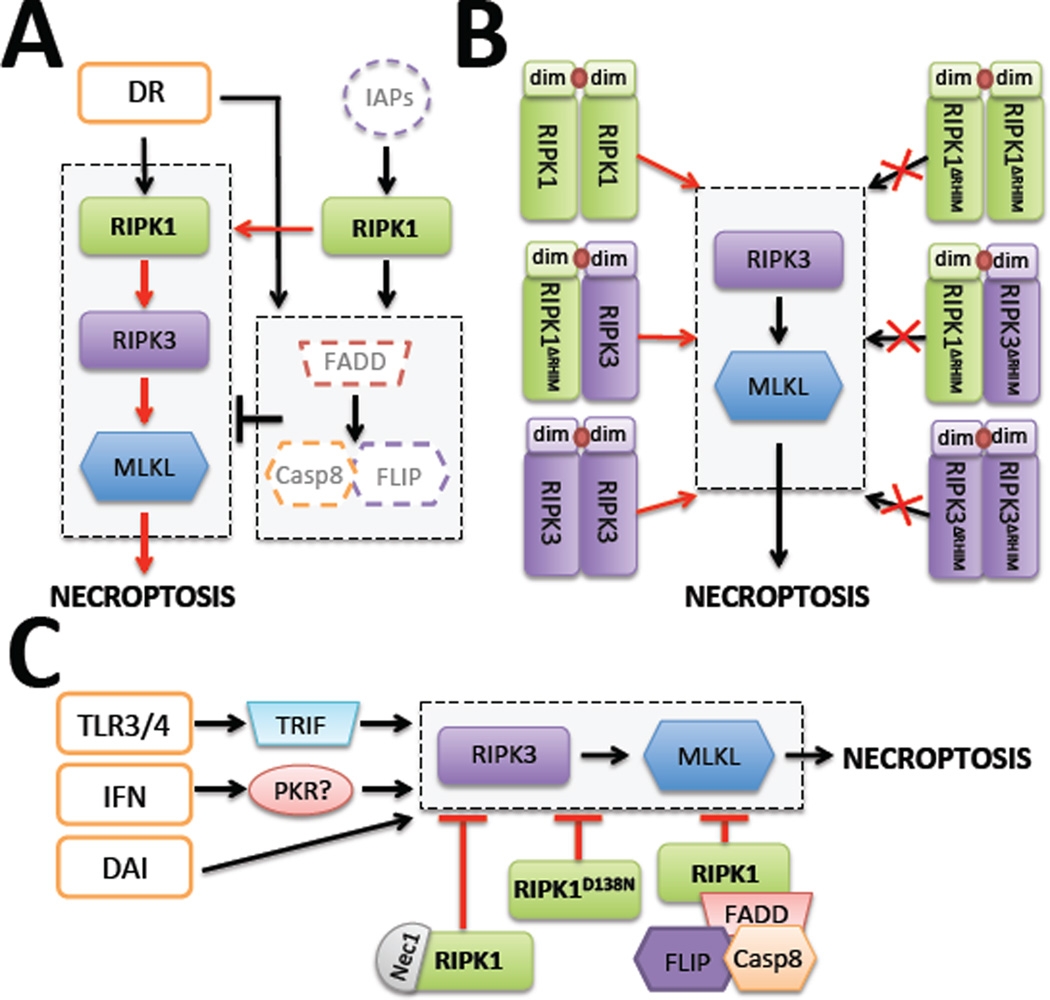
(A) Ligation of death receptors or loss of IAPs induce necroptosis by the formation of a complex containing RIPK1, RIPK3 and MLKL, under conditions of disruption of the inhibitory complex formed by FADD, Caspase-8 and FLIP. (B) Enforced dimerization of RIPK1 or its dimerization with RIPK3 can induce necroptosis only when it can seed the formation of RIPK3 oligomers. ΔRHIM indicates that the molecule has a defective RHIM and therefore cannot interact via this motif. dim: dimerization domain. (C) Different “signal 2s” (e.g., TLR3/4, DAI, interferons) can induce necroptosis via recruitment of RIPK3 in the absence of RIPK1. RIPK1 blocks necroptosis either through recruitment of the inhibitory complex formed by FADD, caspase-8 and FLIP or when it assumes a kinase inactive conformation (by the mutation D138N or by Nec-1 inhibition).

(A) TRAF2, TRAF6 and the E3 ligases cIAP1 and cIAP2 act in concert to add K63-linked ubiquitin chains (K63-Ub) to RIPK1, as does Pel-1. The LUBAC complex, formed by HOIL-1, HOIP, and Sharpin, ubiquitinates RIPK1 with Met-1-linked ubiquitin chains (Met1-Ub). Both modifications stabilize RIPK1 in complex I, activating the NF-κB pathway and inducing the expression of anti-apoptotic proteins, such as c-FLIP. (B) CYLD and OTULIN are deubiquinating enzymes that remove K63-Ub and linear Met1-Ub linkages from RIPK1, respectively, promoting either apoptosis or necroptosis. (C) RIPK1 modified with K48-linked ubiquitin chains (K48-Ub) is targeted for proteasomal degradation, which sensitizes cells to TNF-induced apoptosis (and possibly signal 2-mediated necroptosis, which is untested). This process is mediated by A20, a ubiquitin-editing enzyme that removes K63-Ub from RIPK1 while adding K48-linked ubiquitin chains, and TRIAD3, a K48-Ub E3 ligase.
References
-
- Balachandran S, Thomas E, Barber GN. A FADD-dependent innate immune mechanism in mammalian cells. Nature. 2004;432:401–405. - PubMed
-
- Belz K, Schoeneberger H, Wehner S, Weigert A, Bönig H, Klingebiel T, Fichtner I, Fulda S. Smac mimetic and glucocorticoids synergize to induce apoptosis in childhood ALL by promoting ripoptosome assembly. Blood. 2014;124:240–250. - PubMed
-
- Berger SB, Kasparcova V, Hoffman S, Swift B, Dare L, Schaeffer M, Capriotti C, Cook M, Finger J, Hughes-Earle A, et al. Cutting Edge: RIP1 kinase activity is dispensable for normal development but is a key regulator of inflammation in SHARPIN-deficient mice. J. Immunol. 2014;192:5476–5480. - PMC - PubMed
-
- Bertrand MJM, Milutinovic S, Dickson KM, Ho WC, Boudreault A, Durkin J, Gillard JW, Jaquith JB, Morris SJ, Barker PA. cIAP1 and cIAP2 Facilitate Cancer Cell Survival by Functioning as E3 Ligases that Promote RIP1 Ubiquitination. Mol. Cell. 2008;30:689–700. - PubMed
Publication types
MeSH terms
Substances
Grants and funding
LinkOut - more resources
Full Text Sources
Other Literature Sources
Molecular Biology Databases
Miscellaneous
