

Giant condylomata acuminata of Buschke and Lowenstein: A peristomal variant
- PMID: 25460461
- PMCID: PMC4275778
- DOI: 10.1016/j.ijscr.2014.10.063
Giant condylomata acuminata of Buschke and Lowenstein: A peristomal variant
Abstract
Introduction: Giant condylomata acuminata (GCA) is a rare, locally invasive tumour that may undergo malignant transformation. It was first described a HPV-induced penile tumour which clinically resembled both a squamous cell carcinoma and condyloma acuminatum, often arising from a pre-existing warty lesion. We describe a case of peri-stomal GCA transformation into invasive squamous cell carcinoma (SCC), which is, to our knowledge, the first report of this in the literature.
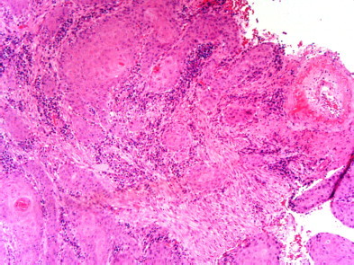
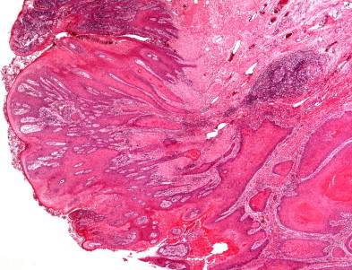
Presentation of case: A 74 year old gentleman developed an acuminate, papillomatous peristomal eruption around a fifty year old ileostomy, with biopsies of the eruption showing reactive changes. Two years later, he developed ulcerating plaques affecting the previously papillomatous areas and an erythematous nodular lesion involving the superior part of the ileostomy and adjacent skin. Histological examination of the ileostomy lesion showed focal small islands of atypical squamous epithelium, and moderately differentiated invasive squamous cell carcinoma was shown in the excised tissue subsequently. Human papillomavirus (HPV type 16), p16 and p53 tumour suppressors were positive in the peri-stomal skin sample.
Discussion and conclusions: Recurring, changing papillomatous lesions in the peristomal area should be reviewed with a high index of suspicion in relation to GCA tumours as they can progress to invasive squamous cell carcinomas.
Keywords: Buschke–Lowenstein tumour; Giant condylomata acuminata; Human papillomavirus; Stoma.
Copyright © 2014 The Authors. Published by Elsevier Ltd.. All rights reserved.
Figures


References
-
- Al-Niaimi F., Lyon C.C. Primary adenocarcinoma in peristomal skin: a case study. Ostomy Wound Manag. 2010;56(1):45–47. - PubMed
-
- Wilson M.S., Scott N.A. Case report – adenocarcinoma arising in an ileostomy managed by resection and an ileo-anal J pouch. Colorectal Dis. 2000;2:185–186. - PubMed
-
- O’Connell P.R., Dozois R.R., Irons G.B., Scheithauer B.W. Squamous cell carcinoma occurring in a skin-grafted ileostomy stoma. Report of a case. Dis Colon Rectum. 1987;30(6):475–478. - PubMed
LinkOut - more resources
Full Text Sources
Other Literature Sources
Research Materials
Miscellaneous
