

Primary intrahepatic malignant epithelioid mesothelioma
- PMID: 25460485
- PMCID: PMC4275834
- DOI: 10.1016/j.ijscr.2014.11.028
Primary intrahepatic malignant epithelioid mesothelioma
Abstract
Introduction: Primary malignant hepatic mesotheliomas are extremely rare. We report the case of a patient with primary intrahepatic malignant mesothelioma who was treated in our department.
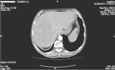
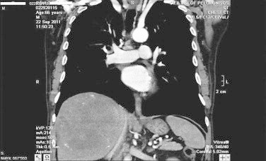
Presentation of case: A 66-year old male patient was admitted to our department for the evaluation of anemia. An abdominal computed tomography scan revealed a large space occupying lesion in the right liver lobe.
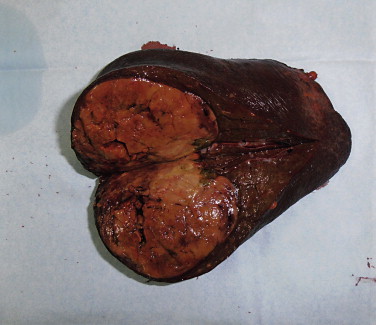
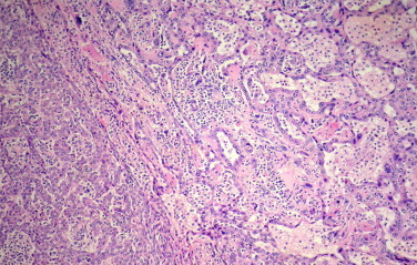
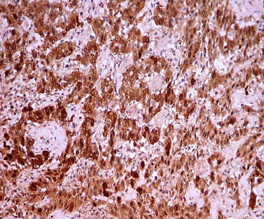
Discussion: The tumor was subsequently resected and a diagnosis of primary intrahepatic malignant mesothelioma was made after pathologic examination. The patient did not receive adjuvant therapy and is currently alive and free of disease, 36 months after the resection.
Conclusion: To our knowledge this is the eighth adult case of primary intrahepatic malignant mesothelioma reported in the literature. These tumors are rarely diagnosed preoperatively. Absence of previous asbestos exposure does not exclude malignant mesothelioma from the differential diagnosis. Proper surgical treatment may offer prolonged survival to the patient, without adjuvant therapy.
Keywords: Epithelioid type; Intrahepatic; Malignant mesothelioma.
Copyright © 2014. Published by Elsevier Ltd.
Figures
References
-
- Teta M.J., Mink P.J., Lau E., Sceurman B.K., Foster E.D. US mesothelioma patterns 1973–2002: indicators of change and insights into background rates. Eur J Cancer Prev. 2008;17:525–534. - PubMed
-
- Imura J., Ichikawa K., Takeda J., Iwasaki Y., Tomita S., Kubota K. Localized malignant mesothelioma of the epithelial type occurring as a primary hepatic neoplasm: a case report with review of the literature. APMIS. 2002;110:789–794. - PubMed
-
- Leonardou P., Semelka R.C., Kanematsu M., Braga L., Woosley J.T. Primary malignant mesothelioma of the liver: MR imaging findings. Magn Reson Imaging. 2003;21:1091–1093. - PubMed
-
- Gutgemann I., Standop J., Fischer H.P. Primary intrahepatic malignant mesothelioma of epithelioid type. Virchows Arch. 2006;448:655–658. - PubMed
LinkOut - more resources
Full Text Sources
Other Literature Sources
Research Materials
