Review
. 2014 Dec:41:183-92.
doi: 10.1016/j.yebeh.2014.09.015. Epub 2014 Oct 26.

Proceedings of the Fifth International Workshop on Advances in Electrocorticography

Affiliations
Review

Proceedings of the Fifth International Workshop on Advances in Electrocorticography

Anthony Ritaccio et al. Epilepsy Behav. 2014 Dec.

Abstract

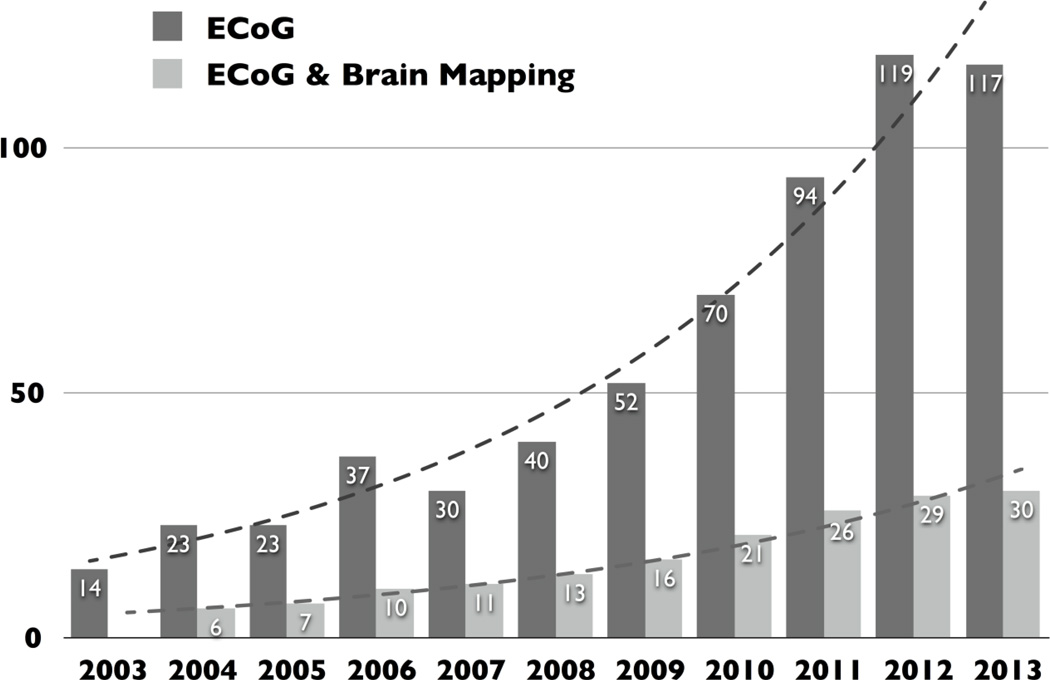
The Fifth International Workshop on Advances in Electrocorticography convened in San Diego, CA, on November 7-8, 2013. Advancements in methodology, implementation, and commercialization across both research and in the interval year since the last workshop were the focus of the gathering. Electrocorticography (ECoG) is now firmly established as a preferred signal source for advanced research in functional, cognitive, and neuroprosthetic domains. Published output in ECoG fields has increased tenfold in the past decade. These proceedings attempt to summarize the state of the art.

Keywords: Brain mapping; Brain–computer interface; Electrical stimulation mapping; Electrocorticography; Functional mapping; Gamma-frequency electroencephalography; High-frequency oscillations; Neuroprosthetics; Seizure detection; Subdural grid.


Conflict of interest statement

Conflict of Interest

We confirm that there are no known conflicts of interest associated with this publication and that there has been no significant financial support for this work that could have influenced its outcome.

Figures

Fig. 1
Fig. 1. Results of simple searches in PubMed for the terms “electrocorticography” and, separately, “electrocorticography and brain mapping”
Results demonstrate a roughly tenfold increase in publication output and investigators.
Fig. 2
Fig. 2. Experiments by Giovanni Aldini (1762–1834) with electrical stimulation of cadavers using voltaic piles (1802)
Fig. 3
Fig. 3. Pairwise 12–20 Hz phase coherence with a pericentral motor cortex
A seed for the coherence measure is chosen by selecting the most index finger-specific site (identified by magnitude of broadband shift), denoted with “ship wheel” symbol. (A) The pairwise phase coherence between sites is calculated with the remainder of the array (during rest periods). The magnitude of the phase coherence (max = 0.32) is reflected in the strength of the color and the electrode diameter, whereas the relative phase-lag of the phase coherence is denoted by the color. (B) To more clearly isolate spatial changes in phase, the complex phase coherence at each site was projected onto the phase of the site of maximum absolute phase coherence. (C) The mean projected phase coherence from each region (shown color coded on inset cortical rendering) is quantified, with error bars denoting the standard error of the mean within each region. (D and E) As in A and B; maximum phase coherence noted in between cortical renderings. (F) Pooled data from subjects 1–9, showing that the 12–20 Hz pairwise phase coherence is conserved within dorsal pericentral cortex, bounded by the precentral sulcus anteriorly and the postcentral sulcus posteriorly. Note that the “anti-phase coherence” is most strongly due to introduced phase coherence in the common-average process (π out of phase) in the electrodes that do not otherwise have a large β-rhythm. See Miller et al. [42] for a complete description.
Fig. 4
Fig. 4. Comparisons between distributions of HGAs and BOLD responses on a template brain
(A) All ECoG electrodes displayed on a template brain. The electrodes (green dots) on the template brain widely covered the lateral aspect of the left frontal and temporal lobes. (B) ECoG electrodes with significant HGA. Different colors of the electrodes indicate individual patients. The electrodes were mainly clustered on the inferior frontal, superior and middle temporal gyri, and precentral gyrus (premotor cortex and face-motor area). (C) A three-dimensional t-map of BOLD response across individual displayed on a template brain. The BOLD responses were widely observed in the frontal lobe, which involves the inferior frontal and precentral gyri. There were additional activated areas in the inferior temporal gyrus, which was sparsely covered by ECoG electrodes. Time-frequency analysis was applied to compare their spatiotemporal profiles.
Fig. 5
Fig. 5. High gamma activity (HGA) vs. blood oxygen level-dependent (BOLD) imaging signal change
Comparison of normalized HGA and BOLD signal show a positive correlation (R = 0.57, p = 0.0002) between these two assessment tools.
Fig. 6
Fig. 6. Temporal dynamics of high gamma activity of noticeable electrode clusters in frontal and temporal lobes
Each line color corresponds to electrode color. X and Y axes indicate latency and high gamma broadband index (HGBI), respectively. HGBI in the frontal lobe lasts longer beyond 1000 msec. On the other hand, rapid decline of HGBI was observed after 500 msec in the temporal lobe.
Fig. 7
Fig. 7. Electrocorticography reveals the neural basis of memories
(A) The amplitude of high gamma (HG; 65–150 Hz) ECoG activity at a site in one patient’s ventral temporal lobe in a working memory task (Modified from Jacobs and Kahana 2009). Shading at top indicates time points that exhibited significant differences in amplitude according to the identity of the viewed item. (B) The amplitude of HG activity for all letters viewed in the task (same electrode as panel A.) (C) Brain image from a patient who spontaneously remembered memories of high school (HS) after stimulation at the indicated electrode (red) in his left ventral temporal lobe. (D) The amplitude of HG activity observed from this electrode when the patient performed a memory task where he remembered HS and non-HS information (same electrode as panel C.)

References

    1. Carey B. Probing brain's depth, trying to aid memory. The New York Times Online; Jul 9, 2014. [Accessed 25 July 2014]. http://www.nytimes.com/2014/07/09/health/probing-brains-depth-trying-to-....
    1. Satel S, Lilienfeld SO. Brainwashed: the Seductive Appeal of Mindless Neuroscience. New York: Basic Books; 2013.
    1. Asano E, Juhász C, Shah A, Sood S, Chugani HT. Role of subdural electrocorticography in prediction of long-term seizure outcome in epilepsy surgery. Brain. 2009;132:1038–1047. - PMC - PubMed
    1. Palmini A, Gambardella A, Andermann F, Dubeau F, da Costa JC, Olivier A, et al. Intrinsic epileptogenicity of human dysplastic cortex as suggested by corticography and surgical results. Ann Neurol. 1995;37:476–487. - PubMed
    1. Wong CH, Birkett J, Byth K, Dexter M, Somerville E, Gill D, et al. Risk factors for complications during intracranial electrode recording in presurgical evaluation of drug resistant partial epilepsy. Acta Neurochir (Wien) 2009;151:37–50. - PubMed

Publication types

MeSH terms