. 2015 Feb:74:114-25.
doi: 10.1016/j.nbd.2014.10.024. Epub 2014 Nov 4.

Hematogenous macrophage depletion reduces the fibrotic scar and increases axonal growth after spinal cord injury

Affiliations

Hematogenous macrophage depletion reduces the fibrotic scar and increases axonal growth after spinal cord injury

Y Zhu et al. Neurobiol Dis. 2015 Feb.

Abstract

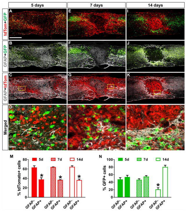
Spinal cord injury (SCI) leads to formation of a fibrotic scar that is inhibitory to axon regeneration. Recent evidence indicates that the fibrotic scar is formed by perivascular fibroblasts, but the mechanism by which they are recruited to the injury site is unknown. Using bone marrow transplantation in mouse model of spinal cord injury, we show that fibroblasts in the fibrotic scar are associated with hematogenous macrophages rather than microglia, which are limited to the surrounding astroglial scar. Depletion of hematogenous macrophages results in reduced fibroblast density and basal lamina formation that is associated with increased axonal growth in the fibrotic scar. Cytokine gene expression analysis after macrophage depletion indicates that decreased Tnfsf8, Tnfsf13 (tumor necrosis factor superfamily members) and increased BMP1-7 (bone morphogenetic proteins) expression may serve as anti-fibrotic mechanisms. Our study demonstrates that hematogenous macrophages are necessary for fibrotic scar formation and macrophage depletion results in changes in multiple cytokines that make the injury site less fibrotic and more conducive to axonal growth.

Keywords: Axonal growth; Bone morphogenetic protein; Fibroblasts; Fibrotic scar; Hematogenous macrophages; Spinal cord injury; Tumor necrosis factor.


Figures

Figure 1
Figure 1. Fibroblasts and macrophages display similar spatiotemporal distribution profile
In the uninjured spinal cord (A–D), Col1α1GFP fibroblasts are around blood vessels while CD11b+ cells represent microglia. At 3 days after injury (EH), fibroblasts are not present in significant numbers (E) and round CD11b+ cells occupy mostly the peripheral edges of the injury site (F). At 7 days (I–L), fibroblasts and CD11b+ macrophages densely populate the injury site. At 14 days (M–P), when the fibrotic scar has matured, it is characterized by a dense population of mostly fibroblasts and CD11b+ macrophages. Similar expression pattern is found at more chronic time points (Q–X). Region of interest (ROI) represents boxed region in the left. n=5 per group. Scale bar in A = 500 μm; D = 50 μm.
Figure 2
Figure 2. Fibrotic scar is occupied by hematogenous macrophages
lysMtdTom>Cx3cr1GFP chimeric mice in which hematogenous macrophages are labeled with tdTomato and microglia are labeled with GFP show that at 5 (A–D) and 7 (E–H) days after SCI, both cell types are interspersed throughout the fibrotic (GFAP area) and astroglial (GFAP+ area) scar. However, at 14 days (I–L), the fibrotic scar is occupied almost entirely by lysMtdTom cells whereas Cx3cr1GFP cells are present mostly in the surrounding astroglial scar region. Majority of lysMtdTom cells are present in the fibrotic scar (GFAP region) at 5, 7 and 14 days after SCI (M, n=3), while the presence of Cx3cr1GFP cells preferentially in the GFAP+ region appears at 14 days but not earlier 29 after injury (N, n=3). *p<0.05 compared to GFAP region in M, compared to 14d GFAP+ region in N using one way ANOVA with Tukey’s post-test. Error bars are s.e.m.
Figure 3
Figure 3. Hematogenous macrophage depletion reduces the fibrotic scar
Treatment with clodronate liposomes significantly reduced the amount of macrophages at both 7 (A, I, Q, n=6 for PBS, n=5 for CLO) and 14 (C, K, S, n=6 for PBS, n=7 for CLO) days after SCI. This reduction in macrophages was associated with a reduction in fibroblasts (B, J, D, L, R, T). Notably, the dense peripheral rim of fibroblasts bordering the astroglial scar was visibly diminished in clodronate treated animas (D, L, F, G, N, O). This reduction in peripheral rim fibroblasts was associated with diminished basal lamina formation between fibroblasts and astrocytes (H, P). The reduction in macrophages and fibroblasts led to a significant increase in the GFAP area at 14 days (E, M, U). Boxed region in D, E is represented by F, G. Boxed region in L, M is represented by N, O. Q, R, S, T were calculated based on percent of GFAP area. *p<0.05 compared to PBS using two-tailed student’s t-test. PBS = PBS liposome. CLO = Clodronate liposome. Scale bar in A–E, I–M, H–P = 500 μm; in F, G, N, O = 100 μm; Error bars are s.e.m.
Figure 4
Figure 4. Liposomes specifically target hematogenous macrophages
At both 7 (A, B, E, F, n=3) and 14 (C, D, G, H, n=3) days after SCI in Col1α1GFP mice, injection of DiI-labeled liposome demonstrates its localization in CD11b+ cells (A–D) and not in Col1α1GFP fibroblasts (E–H). At 14 days after SCI, clodronate treatment in LysMtdTom >Cx3Cr1GFP chimeric mice (I–N, n=3 for clodronate, n=4 for PBS) shows a visible reduction in hMΦ (LysMtdTom) but not microglia (Cx3Cr1GFP). B, D, F, H are single planes from a confocal z-stack series and represent boxed region in A, C, E, G respectively. Scale bar in A, I = 500 μm; B = 50 μm.
Figure 5
Figure 5. Fibroblast reduction is not due to reduced angiogenesis after macrophage depletion
At 14 days after SCI, clodronate treatment (n=5, D–F) resulted in a significant decrease in both fibroblast density and blood vessel volume (J, K, Video 3, 4) as compared to PBS liposome treatment (n=4, A–C, J–K, Video 1, 2). While sunitinib treatment (G–I, n=6) also decreased blood vessel volume, it did not significantly affect fibroblast density (L, M, Video 5, 6). A–I are single optical sections from image stacks obtained using light sheet fluorescent microscopy (Video 1, 3, 5). Vehicle in the clodronate study was PBS liposome (A–C, J–K). Vehicle in the sunitinib study was 2.5% DMSO (n=4, images not shown). *p<0.05 compared to vehicle using two-tailed student’s t-test. Scale bar = 300 μm in A–I. Error bars are s.e.m.
Figure 6
Figure 6. Fibrotic scar reduction is associated with increased growth of neurofilament+ axons
At 14 days after SCI, the number of neurofilament+ axons in the fibrotic scar (GFAP region) was similar between PBS (A–C, n=3) and clodronate liposome (D–F, n=4) treated animals (M). However, at 56 days after SCI, the number of neurofilament+ axons in the clodronate liposome treated animals (J–L, M, n=7) was significantly higher than PBS liposome treated controls (G–I, M, n=7). The GFAP area was smaller in clodronate liposome treated animals at 56 days after injury (N). Region of interest (ROI) represents boxed region to the left. *p<0.05 compared to the other groups using two way ANOVA with Tukey’s post-test in M or two tailed student’s t-test in N. Scale bar in A = 500 μm; in C = 50 μm. PBS = PBS liposome. CLO = Clodronate liposome. Error bars are s.e.m.
Figure 7
Figure 7. Macrophage depletion alters both pro- and anti-fibrotic cytokine expression
A) Volcano plot of all 84 cytokines tested in the PCR array comparing the injury sites of clodronate and PBS treated mice at 7 days after injury (n=5 per group). Dotted vertical line represents 1.5 fold change and solid horizontal line represents p=0.05 (unpaired t-test comparing clodronate and PBS treated mice for each gene). Green squares are BMPs and magenta triangles are Tnfsf. B) Bar graph showing fold change in differentially expressed genes between clodronate and PBS treated mice (each bar represents average of clodronate (n=5)/average of PBS (n=5)).

References

    1. Arenkiel BR, et al. Activity-induced remodeling of olfactory bulb microcircuits revealed by monosynaptic tracing. PloS one. 2011;6:e29423. - PMC - PubMed
    1. Ashbaugh JJ, et al. IL7Ralpha contributes to experimental autoimmune encephalomyelitis through altered T cell responses and nonhematopoietic cell lineages. Journal of immunology. 2013;190:4525–34. - PMC - PubMed
    1. Basso DM, et al. Basso Mouse Scale for locomotion detects differences in recovery after spinal cord injury in five common mouse strains. Journal of neurotrauma. 2006;23:635–59. - PubMed
    1. Becker K, et al. Chemical clearing and dehydration of GFP expressing mouse brains. PloS one. 2012;7:e33916. - PMC - PubMed
    1. Blomster LV, et al. Mobilisation of the splenic monocyte reservoir and peripheral CX(3)CR1 deficiency adversely affects recovery from spinal cord injury. Experimental neurology. 2013;247:226–40. - PubMed

Publication types

MeSH terms

Substances