. 2015 Jan 15:475:88-95.
doi: 10.1016/j.virol.2014.11.008. Epub 2014 Nov 27.

An MHC class I immune evasion gene of Marek׳s disease virus

Affiliations

An MHC class I immune evasion gene of Marek׳s disease virus

Cari Hearn et al. Virology. .

Abstract

Marek׳s disease virus (MDV) is a widespread α-herpesvirus of chickens that causes T cell tumors. Acute, but not latent, MDV infection has previously been shown to lead to downregulation of cell-surface MHC class I (Virology 282:198-205 (2001)), but the gene(s) involved have not been identified. Here we demonstrate that an MDV gene, MDV012, is capable of reducing surface expression of MHC class I on chicken cells. Co-expression of an MHC class I-binding peptide targeted to the endoplasmic reticulum (bypassing the requirement for the TAP peptide transporter) partially rescued MHC class I expression in the presence of MDV012, suggesting that MDV012 is a TAP-blocking MHC class I immune evasion protein. This is the first unique non-mammalian MHC class I immune evasion gene identified, and suggests that α-herpesviruses have conserved this function for at least 100 million years.

Keywords: Immune evasion; Major histocompatibility complex class I; Marek׳s disease virus.


Figures

Figure 1
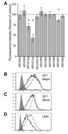
Figure 1. MDV012 reduces cell-surface MHC class I
(A) DF1 cells were transfected with MDV genes as indicated, cloned into pEGFP-N1 (MDV012s) or pTracerCMV2 (all other genes). Two days after transfection the cells were stained for major MHC class I isoform, and transfected cells (GFP positive) were analyzed by flow cytometry. Data are shown as mean fluorescence intensity as a percent of cells transfected with empty vector alone (average of at least three experiments +/− standard deviation). Samples in which the 95% confidence interval does not overlap 100% are indicated with an asterisk (*). (B–D) DF1 (B, C) or LMH (D) chicken cells were transfected with empty vector (thin trace) or with MDV012s (thick trace) and stained for chicken MHC class I using (B) chicken antiserum specific for the major allele of B21, (C) chicken antiserum specific for the minor allele of B21, or (D) a monoclonal antibody specific for chicken MHC class I. Shaded trace: Irrelevant antibody (background staining). Each experiment is representative of at least three replicates.
Figure 2
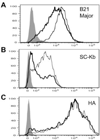
Figure 2. MDV012 reduces expression of H-2Kb, but not of influenza HA, in chicken cells
DF1 cells were transfected with empty vector (thin traces) or with MDV012s (thick traces) and co-transfected with (B) single-chain β2-m/H-2Kb fusion protein (SC-Kb) or (C) with influenza hemagglutinin (HA). After two days, cells were stained with (A) anti-major B21 chicken antiserum, (B) anti-H-2Kb (B8.24.3), or (C) anti- HA (H36.5-4.2). Cells were gated on GFP to limit analysis to transfected cells. Shaded traces: Irrelevant antibodies (background staining). Each experiment is representative of at least three replicates.
Figure 3
Figure 3. B21-binding peptide (R10V) targeted to the ER, but not the cytosol, rescues cell-surface expression of B21 in the presence of MDV012
(A) DF1 cells (B21 +) or LMH cells (B21 -) were transfected with MDV012s (dark bars) or with empty vector as a control (open bars), and co-transfected with either empty vector (“Ctrl”), with the peptide R10V as a cytosolic ubiquitin fusion protein (“R10V”, cytosolic), or with an ER-targeted version of R10V (“ss-R10V”). Two days later, the cells were stained with anti-major B21 chicken serum (A) or with anti-chicken MHC class I (B) and analyzed by flow cytometry.
Figure 4
Figure 4. MDV012 orthologues
MDV012-related proteins of Marek’s Disease Virus (MDV), gallid herpesvirus 3 (GaHV3), Meleagrid herpesvirus 1 (MeHV1), and Duck enteritis virus (DEV). Regions of identity are boxed. The junction between exons 1 and 2 in MDV, GaHV3, and DEV is indicated with an arrow.

References

    1. Atkins KE, Read AF, Savill NJ, Renz KG, Islam AF, Walkden-Brown SW, Woolhouse MEJ. Vaccination and reduced cohort duration can drive virulence evolution: Marek’s disease virus and industrialized agriculture. Evolution. 2013;67:851–860. - PubMed
    1. Bachmair A, Finley D, Varshavsky A. In vivo half-life of a protein is a function of its amino-terminal residue. Science. 1986;234:179–186. - PubMed
    1. Bennett NJ, May JS, Stevenson PG. Gamma-herpesvirus latency requires T cell evasion during episome maintenance. PLoS Biol. 2005;3:e120. - PMC - PubMed
    1. Briles WE, Stone HA, Cole RK. Marek's disease: effects of B histocompatibility alloalleles in resistant and susceptible chicken lines. Science. 1977;195:193–195. - PubMed
    1. Burgos JS, Serrano-Saiz E, Sastre I, Valdivieso F. ICP47 mediates viral neuroinvasiveness by induction of TAP protein following intravenous inoculation of herpes simplex virus type 1 in mice. J. Neurovirol. 2006;12:420–427. - PubMed

Publication types