Review
. 2015 May:77-78:164-71.
doi: 10.1016/j.ymeth.2014.11.008. Epub 2014 Nov 15.

Analysis of intracellular PTEN signaling and secretion

Affiliations
Review

Analysis of intracellular PTEN signaling and secretion

Cindy Hodakoski et al. Methods. 2015 May.

Abstract

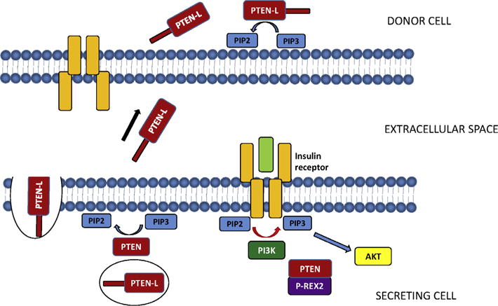
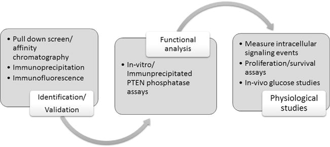
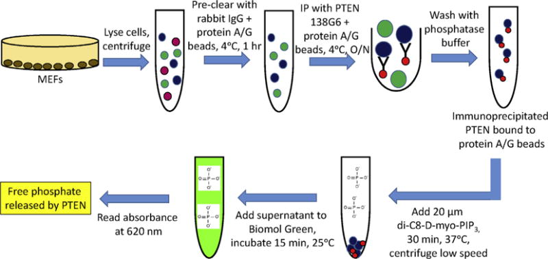
The tumor suppressor PTEN dephosphorylates PIP3 to inhibit PI3K signaling in cells. Altering PTEN intracellular signaling can therefore significantly affect cell behavior. Two novel mechanisms of PTEN regulation including the secretion and entry of the translational variant PTEN-L, and enzymatic inhibition by the interacting protein P-REX2, have been shown to modulate PI3K signaling, cellular proliferation and survival, and glucose metabolism. Here, we review the methods used to identify and validate the existence of both PTEN-L and the P-REX2-PTEN complex, to determine their effects on PTEN phosphatase activity, and to examine their role in cellular physiology.

Keywords: P-REX2; PTEN; PTEN-L; Secretion.


Figures

Fig. 1
Fig. 1
Regulation of intracellular PTEN signaling by PTEN-L and P-REX2. PTEN-L behaves similarly to canonical PTEN, dephosphorylating PIP3 to PIP2. In addition, PTEN-L is also packaged into secretory vesicles and released into the extracellular space. PTEN-L can then enter other cells, increasing the intracellular dose of PTEN in that cell and decreasing PIP3 levels (11). PTEN is also regulated by P-REX2 in response to insulin signaling. P-REX2 binds PTEN and inhibits its phosphatase activity toward PIP3, thus enhancing PI3K signaling in the cell (15, 18).
Fig. 2
Fig. 2
Schematic of methods used to identify PTEN-L and the PTEN-P-REX2 complex, characterize their enzymatic function, and uncover their physiological significance in cells.
Fig. 3
Fig. 3
GST-PTEN pull down screen (15). (A) Generating immobilized GST and GST-tagged PTEN. BL21(DE3) pLysE bacteria transformed with plasmids encoding GST and GST-PTEN are grown to exponential growth and protein expression is induced with 0.1 mM IPTG. After incubation at 16 degrees overnight, cells are sonicated for 40 min in 40 mL lysis buffer and centrifuged at 20,000g for 1 h, and filtered. Glutathione–Sepharose beads are then added for a final concentration of 10 mg per 1 mL glutathione Sepharose. The beads are then washed six times with BC500 buffer followed by two washes with BC200 buffer. (B) GST-PTEN pull-down screen. DBTRG cell lysates are generated as described in Section 2.1. Lysates are pre-cleared by passing them over a column containing GST beads. Pre-cleared lysates are then incubated with either GST-PTEN or GST beads overnight at 4 °C, along with a mock column of GST-PTEN and lysis buffer. The beads are washed 10 times with BC200 buffer wash buffer, and proteins are eluted with hypotonic buffer containing 1 M NaCl. Elutions are resolved by SDS–PAGE and visualized by silver stain, and protein bands specific to the GST-PTEN column are excised and peptide sequenced by MALDI-TOF mass spectrometry.
Fig. 4
Fig. 4
Purification of RFP-tagged PTEN-L (11). (A) Bacterial lysates expressing PTEN-L containing both an RFP and V5/His tag are passed over a HisTrap nickel column. Ni2+ ions form complexes with the histidines present on the His-tagged protein, and unbound protein passes through the column. (B) Proteins bound to the column are eluted with buffer containing 500 mM imidazole, which competes for binding to nickel. (C) The eluted protein containing PTEN-L is passed over a HiTrap heparin column. Heparin binding proteins, including PTEN-L, bind to the column, while unbound proteins are washed away. (D) PTEN-L is eluted with buffer containing 1 M NaCl.
Fig. 5
Fig. 5
Immunoprecipitation PTEN phosphatase assays (18) MEFs are washed in PBS, lysed, and centrifuged at 16,000g for 30 min at 4 °C. Lysates are pre-cleared with 2 μg rabbit IgG plus 40 μL protein A/G beads for 1 h at 4 °C. PTEN is immunoprecipitated with 138G6 antibody plus 40 μL protein A/G beads overnight at 4 °C. Beads are washed with phosphatase buffer and incubated with 20 μM soluble di-C8-D-myo-PIP3 for 30 min at 37 °C. The beads are removed from the reaction mixture by low speed centrifugation, and the supernatant is added to 100 μL Biomol Green reagent for 15 min. The absorbance level at 620 nm is the measure of free phosphate released by PTEN.

References

    1. Maehama T, Dixon JE. J Biol Chem. 1998;273:13375–13378. - PubMed
    1. Lee JO, Yang H, Georgescu MM, Di Cristofano A, Maehama T, Shi Y, et al. Cell. 1999;99:323–334. - PubMed
    1. Leslie NR, Downes CP. J Biochem. 2004;382:1–11. - PMC - PubMed
    1. Stambolic V, Suzuk A, de la Pompa JL, Brothers GM, Mirtsos C, Sasaki T, et al. Cell. 1998;95:29–39. - PubMed
    1. Sun H, Lesche R, Li DM, Liliental J, Zhang H, Gao J, et al. Proc Natl Acad Sci USA. 1999;96:6199–6204. - PMC - PubMed

MeSH terms

LinkOut - more resources