. 2015 Mar;12(1):123-30.
doi: 10.1016/j.pdpdt.2014.10.009. Epub 2014 Nov 13.

Low-level light therapy potentiates NPe6-mediated photodynamic therapy in a human osteosarcoma cell line via increased ATP

Affiliations

Low-level light therapy potentiates NPe6-mediated photodynamic therapy in a human osteosarcoma cell line via increased ATP

Shang-Ru Tsai et al. Photodiagnosis Photodyn Ther. 2015 Mar.

Abstract

Background: Low-level light therapy (LLLT) is used to stimulate healing, reduce pain and inflammation, and preserve tissue from dying. LLLT has been shown to protect cells in culture from dying after various cytotoxic insults, and LLLT is known to increase the cellular ATP content. Previous studies have demonstrated that maintaining a sufficiently high ATP level is necessary for the efficient induction and execution of apoptosis steps after photodynamic therapy (PDT).

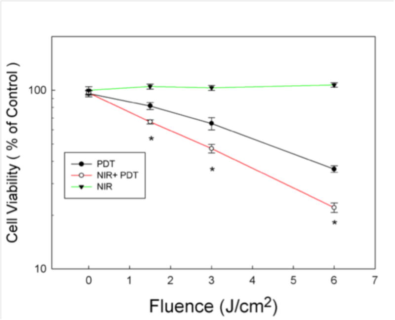
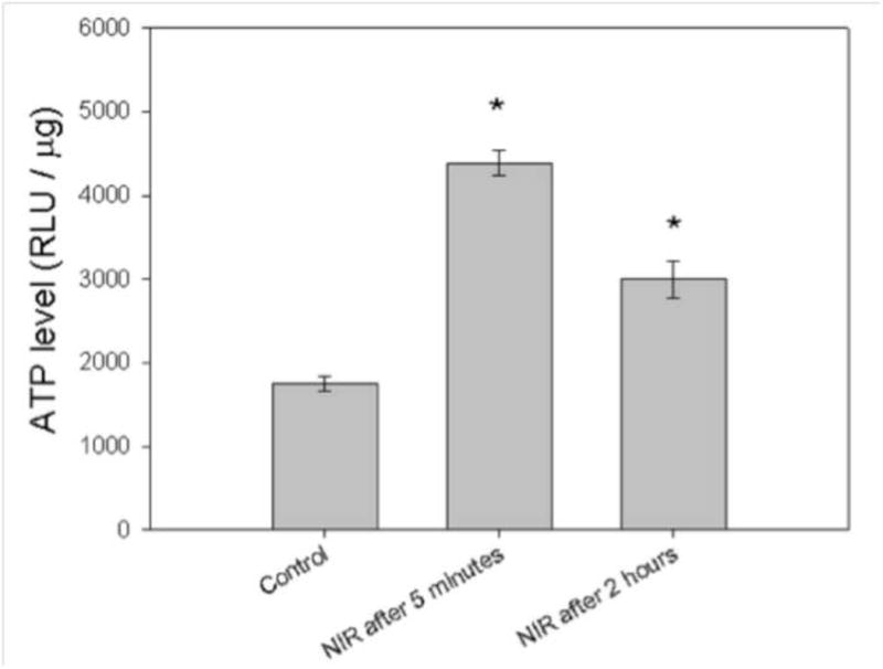
Methods: We asked whether LLLT would protect cells from cytotoxicity due to PDT, or conversely whether LLLT would enhance the efficacy of PDT mediated by mono-l-aspartyl chlorin(e6) (NPe6). Increased ATP could lead to enhanced cell uptake of NPe6 by the energy dependent process of endocytosis, and also to more efficient apoptosis. In this study, human osteosarcoma cell line MG-63 was subjected to 1.5J/cm(2) of 810nm near infrared radiation (NIR) followed by addition of 10μM NPe6 and after 2h incubation by 1.5J/cm(2) of 652nm red light for PDT.

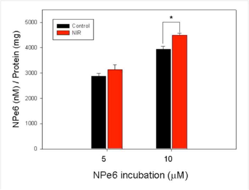
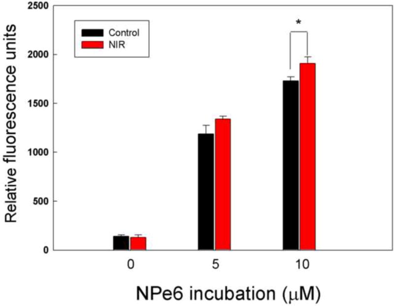
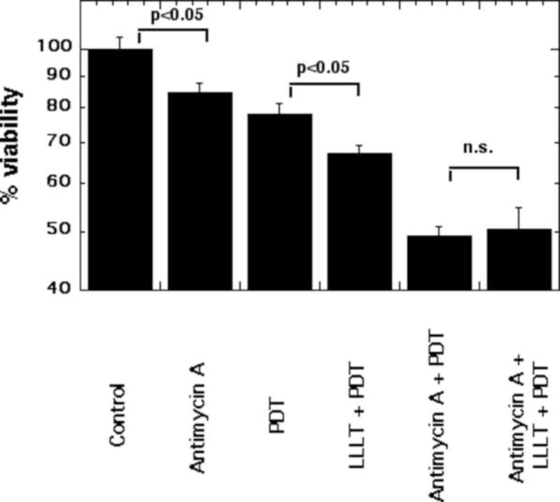
Results: PDT combined with LLLT led to higher cell death and increased intracellular reactive oxygen species compared to PDT alone. The uptake of NPe6 was moderately increased by LLLT, and cellular ATP was increased. The mitochondrial respiratory chain inhibitor antimycin A abrogated the LLLT-induced increase in cytotoxicity.

Conclusions: Taken together, these results demonstrate that LLLT potentiates NPe6-mediated PDT via increased ATP synthesis and is a potentially promising strategy that could be applied in clinical PDT.

Keywords: Adenosine triphosphate; Low level light therapy; Lysosomal uptake; Mono-l-aspartyl chlorin(e6); Photobiomodulation; Photodynamic therapy.


Figures

Figure 1
Figure 1. Cell viability of MG-63 cells pretreated with NIR-LLLT followed by PDT
MG-63 cells were treated with or without NIR-LLLT (fluence 1.5 J/cm2) before 10 μM NPe6 incubation, and then treated with PDT at different fluences 0, 1.5, 3 and 6 J/cm2. (Open circle and closed circle). Additionally, MG-63 cells were treated with NIR-LLLT alone at various fluences 0, 1.5, 3 and 6 J/cm2, cell viability was measured at 24 hours (Closed triangle). Cell viability was measured at 24 hours after PDT or NIR Data are the mean ± SD. An asterisk represents p value < 0.05 as a significant difference between NIR + PDT and PDT alone. All results in this figure are representative of experiments performed in triplicate.
Figure 2
Figure 2. Quantification of NIR enhances cell uptake of NPe6 in MG-63
MG-63 cells were treated with or without 1.5 J/cm2 of 810 nm NIR-LLLT before NPe6 incubation for 2 hours. NPe6 was extracted, corrected for total protein concentration and fluorescence level determined in each cell lysate. Data are the mean ± SD of the fluorescent signal. An asterisk represents the p value < 0.05 as a significant difference between control and NIR-LLLT cells. All results in this figure are representative of experiments performed in triplicate.
Figure 3
Figure 3. NIR-LLLT combined with PDT induced ROS generation determined by flow cytometry
MG-63 cells were pretreated with 1.5 J/cm2 of 810 nm NIR-LLLT and incubated with NPe6 for 2 h, exposed to 1.5 J/cm2 652 nm red light, and then stained with H2DCFDA for 30 minutes to measure the level of ROS by flow cytometry. Data are the mean ± SD of the fluorescent signal. An asterisk represents the p value < 0.05 as a significant difference between control and NIR-treated cells. All results in this figure are representative of experiments performed in triplicate.
Figure 4
Figure 4. Subcellular localization of the NPe6 in MG-63 cells
Confocal fluorescence images of subcellular localization of the MG-63 cells with or without 1.5 J/cm2 of 810 nm NIR-LLLT and 2 h of incubation with 10 μM NPe6. Cells were stained with organelle-specific fluorescent probes. NPe6 is red, mitochondria are green, lysosomes are yellow, and nucleus is blue, respectively. Scale bar indicates 10 μm.
Figure 5
Figure 5. NIR-LLLT stimulates ATP synthesis in MG-63 cells
MG-63 cells were treated with 1.5 J/cm2 of 810 nm NIR-LLLT. After 5 min and 2 h, cells were lysed and Cell-Titer Glo Assay mix used to determine ATP levels. Data are the mean ± SD. An asterisk represents the p value < 0.05 as a significant difference between control and NIR-treated cells. All results in this figure are representative of experiments performed in triplicate.
Figure 6
Figure 6. NIR-LLLT potentiation of NPe6-PDT is abrogated by antimycin A
After 1 hour incubation with 1 nM antimycin A, MG-63 cells were treated with or without 1.5 J/cm2 of 810 nm NIR-LLLT before 2 hour NPe6 incubation, and then exposed to 1.5 J/cm2 652 nm red light for PDT. Cell viability was measured at 24 h after PDT. Data are the mean ± SD of the fluorescent signal. p value < 0.05 showed a significant difference between the indicated groups. All results in this figure are representative of experiments performed in triplicate.

References

    1. Dolmans DEJGJ, Fukumura D, Jain RK. Photodynamic therapy for cancer. Nat Rev Cancer. 2003;3:380–7. - PubMed
    1. Allison RR, Moghissi K. Photodynamic Therapy (PDT): PDT Mechanisms. Clinical endoscopy. 2013;46:24–9. - PMC - PubMed
    1. Castano AP, Demidova TN, Hamblin MR. Mechanisms in photodynamic therapy: part one—photosensitizers, photochemistry and cellular localization. Photodiagnosis and Photodynamic Therapy. 2004;1:279–93. - PMC - PubMed
    1. Rosenkranz AA, Jans DA, Sobolev AS. Targeted intracellular delivery of photosensitizers to enhance photodynamic efficiency. Immunol Cell Biol. 2000;78:452–64. - PubMed
    1. Anselmo AC, Mitragotri S. An Overview of Clinical and Commercial Impact of Drug Delivery Systems. Journal of controlled release: official journal of the Controlled Release Society. 2014 - PMC - PubMed

Publication types

MeSH terms