. 2015 Feb 12:286:87-96.
doi: 10.1016/j.neuroscience.2014.11.030. Epub 2014 Nov 26.

Zebrafish calsyntenins mediate homophilic adhesion through their amino-terminal cadherin repeats

Affiliations

Zebrafish calsyntenins mediate homophilic adhesion through their amino-terminal cadherin repeats

H Ortiz-Medina et al. Neuroscience. .

Abstract

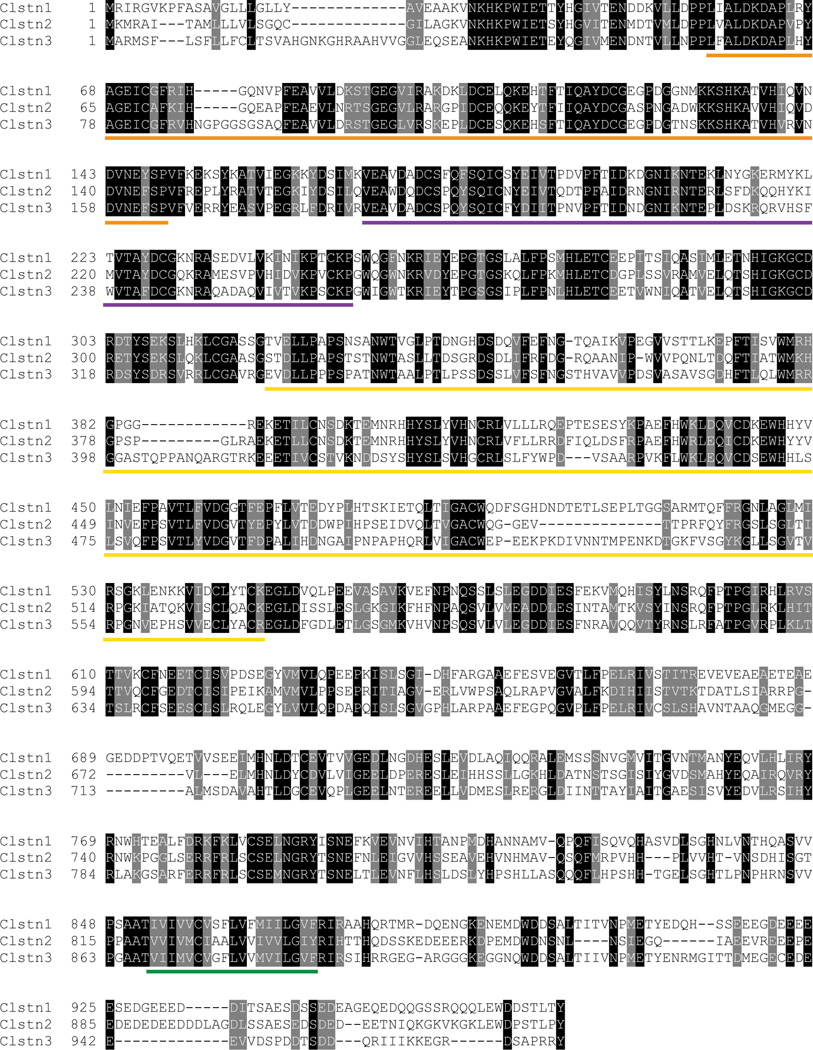
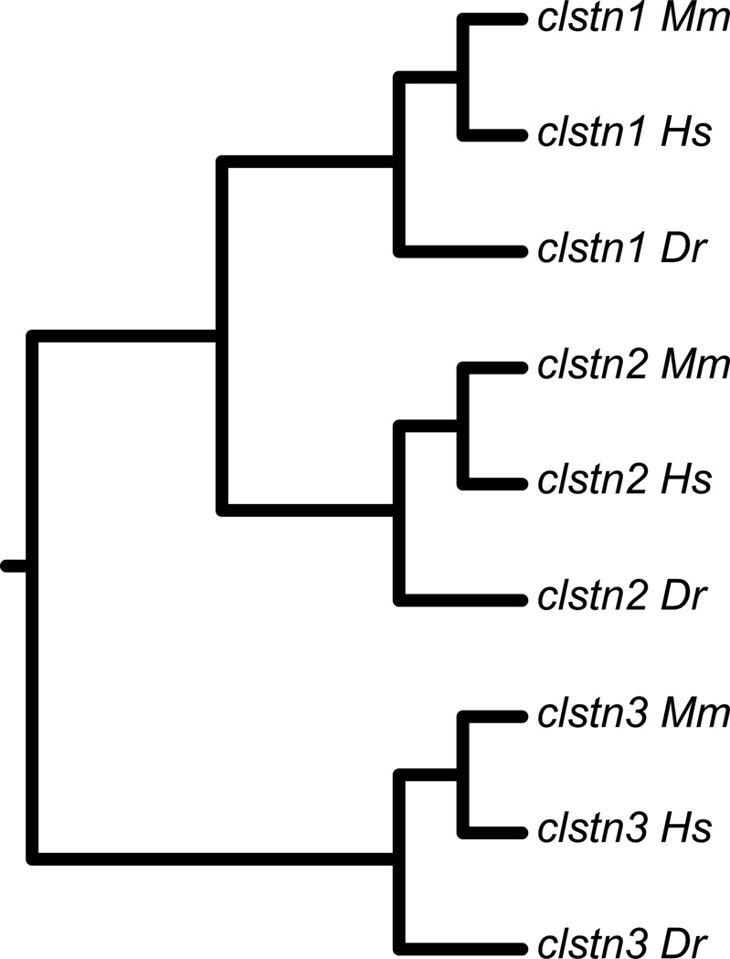
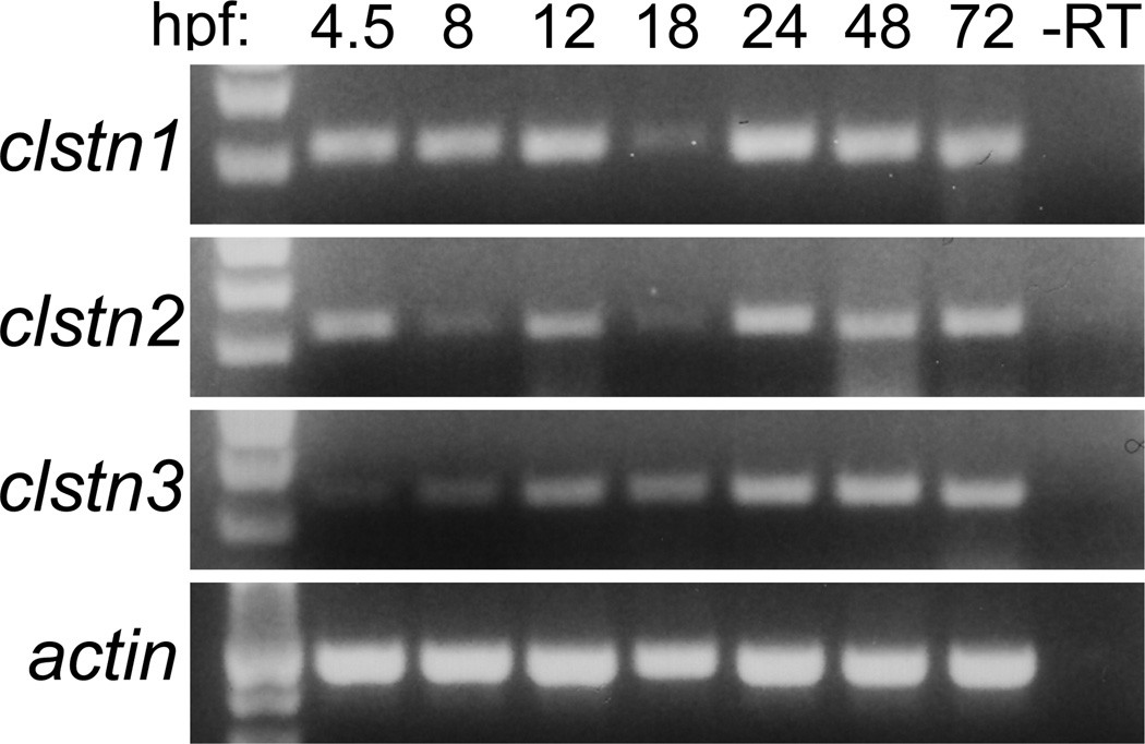
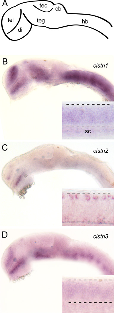
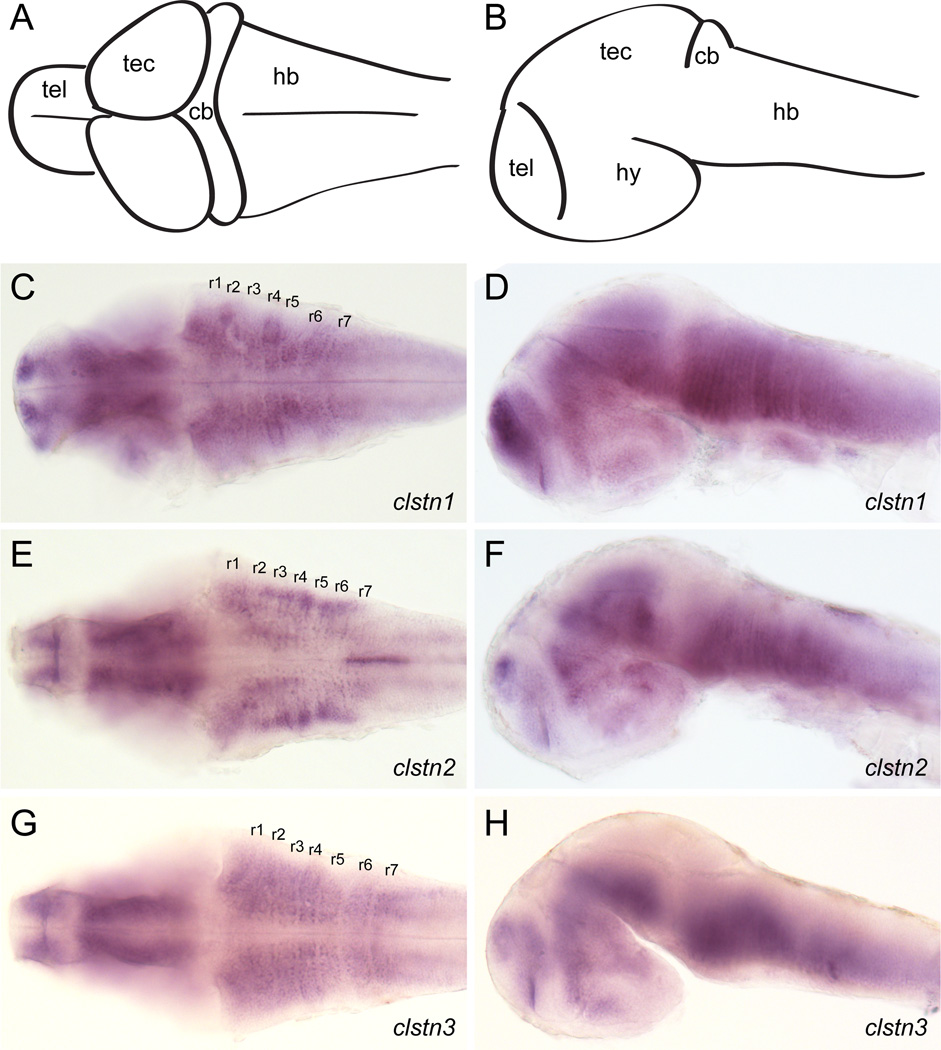
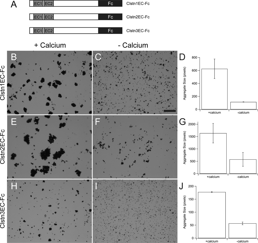
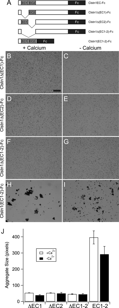
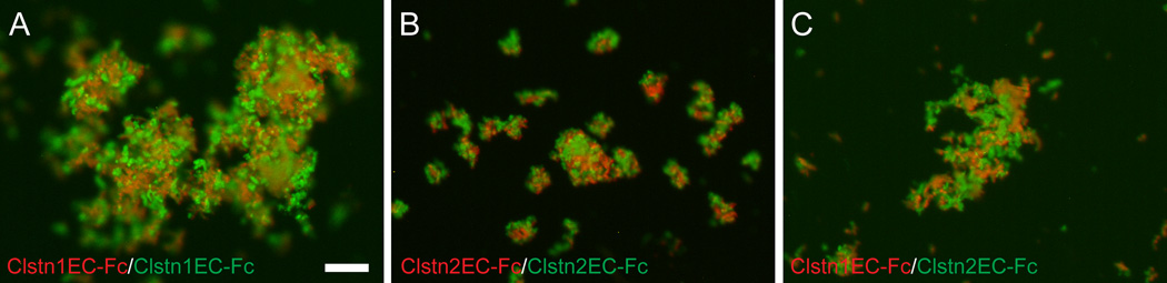
The calsyntenins are atypical members of the cadherin superfamily that have been implicated in learning in Caenorhabditis elegans and memory formation in humans. As members of the cadherin superfamily, they could mediate cell-cell adhesion, although their adhesive properties have not been investigated. As an initial step in characterizing the calsyntenins, we have cloned clstn1, clstn2 and clstn3 from the zebrafish and determined their expression in the developing zebrafish nervous system. The three genes each have broad, yet distinct, expression patterns in the zebrafish brain. Each of the ectodomains mediates homophilic interactions through two, amino-terminal cadherin repeats. In bead sorting assays, the calsyntenin ectodomains do not exhibit homophilic preferences. These data support the idea that calsyntenins could either act as adhesion molecules or as diffusible, homophilic or heterophilic ligands in the vertebrate nervous system.

Keywords: adhesion; cadherin; calsyntenin; zebrafish.


Figures

Figure 1
Figure 1. Protein sequences and alignments for the zebrafish calsyntenins
The three calsyntenin proteins exhibit two, amino-terminal cadherin repeats (orange and purple lines), an LGN domain, (yellow line), a single pass transmembrane segment (green line) and a short cytoplasmic tail.
Figure 2
Figure 2. Phylogenetic analysis of zebrafish Calsyntenins
The protein sequences of the zebrafish calsyntenins were aligned with those of human and mouse, then analyzed by MAFFT/ClustalX. Each of the zebrafish calsyntenin protein sequences clusters with a human and mouse pair, indicating that the zebrafish genes encode homologs for Clstn1, Clstn2 and Clstn3.
Figure 3
Figure 3. Temporal expression of calsyntenins during zebrafish development
RT-PCR was performed using cDNA prepared from 4.5 hpf to 72 hpf embryos. Expression was evident as early as 4.5 hpf and continued throughout all developmental stages analyzed. Actin was used as a control.
Figure 4
Figure 4. Expression of calsyntenins in 1 day old zebrafish embryos
A. Diagram of a lateral view of a 1 day old zebrafish brain. Abbreviations: tel, telencephalon; di, diencephalon; teg, tegmentum; tec, optic tectum; cb, cerebellum; hb, hindbrain. B–D. Expression of clstn1 (B), clstn2 (C), and clstn3 (D) in the brain and spinal cord of 1 day zebrafish embryos, as determined by in situ hybridization (inset; sc, spinal cord).
Figure 5
Figure 5. Expression of calsyntenins in 2 day old zebrafish embryos
A, B. Diagrams of a lateral view (A) and dorsal view (B) of a 2 day old zebrafish brain. Abbreviations: tel, telencephalon; hy, hypothalamus; tec, optic tectum; cb, cerebellum; hb, hindbrain. C, D. Expression of clstn1 in the brain of 2 day zebrafish embryos, as determined by in situ hybridization. Expression is evident in all the major subdivisions of the brain and is particularly strong in the telencephalon, as well as showing a ladder-like expression in the hindbrain. Rhombomeres are labeled r1–r7. E, F. Expression of clstn2 in the brain of 2 day zebrafish embryos, as determined by in situ hybridization. Like clstn1, expression is evident in all the major subdivisions of the brain. Segmental labeling of lateral cell clusters is apparent in the hindbrain. G, H. Expression of clstn3 in the brain 2 day zebrafish embryos, as determined by in situ hybridization. The overall pattern of expression is comparable to the other calsyntenins, though clstn3 appears to be absent from the optic tectum.
Figure 6
Figure 6. Calsyntenin ectodomains mediate bead aggregation
A. The ectodomain of each calsyntenin was fused to the Fc portion of human IgG and the CstnEC-Fc fusions were used in bead aggregation assays. B–H. Each of the three Calsyntenin ectodomains, Clstn1EC-Fc (B, C, D), Clstn2EC-Fc (E, F, G) and Clstn3EC-Fc (H, I, J), are able to mediate calcium-dependent bead aggregation (n=3 for each condition). The aggregates formed by Clstn3EC-Fc (F) were smaller than those formed by either Clstn1EC-Fc (B) or Clstn2EC-Fc (D). Each fusion-protein also exhibits some calcium-independent aggregation (C, E, G). In each case, the calcium-dependent increase was statistically significant (p<0.05, Student's T-test). Scale bar = 50 µm.
Figure 7
Figure 7. Cadherin repeats are required for bead aggregation by Calsyntenin-1
A. A series of domain deletions of the Clstn1EC-Fc were generated to test the role of the cadherin repeats, EC1 and EC2. B–G. Deletion of EC1 (B,C), EC2 (D,E) or EC1–EC2 (F,G) abolish calcium-dependent and calcium-independent bead aggregation. Scale bar = 50 µm. H, I. The cadherin repeats EC1–EC2 are sufficient to mediate calcium-dependent bead aggregation. There is also an increase in calcium-independent bead aggregation. J. Deletion of either cadherin repeat is sufficient to abolish bead aggregation, while the two cadherin repeats are sufficient for bead aggregation (n=3 for each condition). The two cadherin repeats exhibit only a modest, but significant, calcium-dependence (p=0.04, Student's T-test).
Figure 8
Figure 8. Heterophilic interactions by calsyntenin ectodomains
A. The ectodomain of Clstn1 (Clstn1EC-Fc) was used to coat green or red fluorescent beads. Coated beads formed mixed aggregates. Scale bar = 20 µm. B. As for Clstn1EC-Fc (A), green and red beads coated with Clstn2EC-Fc intermixed. C. Red fluorescent beads were coated with Clstn1EC-Fc and green fluorescent beads were coated with Clstn2EC-Fc. When allowed to aggregate in the presence of calcium, the green and red beads intermixed, as in A and B.

Similar articles

Cited by

References

    1. Araki Y, Kawano T, Taru H, Saito Y, Wada S, Miyamoto K, Kobayashi H, Ishikawa HO, Ohsugi Y, Yamamoto T, Matsuno K, Kinjo M, Suzuki T. The novel cargo Alcadein induces vesicle association of kinesin-1 motor components and activates axonal transport. EMBO J. 2007;26:1475–1486. - PMC - PubMed
    1. Araki Y, Miyagi N, Kato N, Yoshida T, Wada S, Nishimura M, Komano H, Yamamoto T, De Strooper B, Yamamoto K, Suzuki T. Coordinated metabolism of Alcadein and amyloid beta-protein precursor regulates FE65-dependent gene transactivation. J Biol Chem. 2004;279:24343–24354. - PubMed
    1. Araki Y, Tomita S, Yamaguchi H, Miyagi N, Sumioka A, Kirino Y, Suzuki T. Novel cadherin-related membrane proteins, Alcadeins, enhance the X11-like protein-mediated stabilization of amyloid beta-protein precursor metabolism. J Biol Chem. 2003;278:49448–49458. - PubMed
    1. Biswas S, Emond MR, Jontes JD. Protocadherin-19 and N-cadherin interact to control cell movements during anterior neurulation. J Cell Biol. 2010;191:1029–1041. - PMC - PubMed
    1. Blevins CJ, Emond MR, Biswas S, Jontes JD. Differential expression, alternative splicing, and adhesive properties of the zebrafish delta1-protocadherins. Neuroscience. 2011;199:523–534. - PubMed

Publication types