. 2014 Dec 2;9(12):e114198.
doi: 10.1371/journal.pone.0114198. eCollection 2014.

Osteoblast-specific deletion of Pkd2 leads to low-turnover osteopenia and reduced bone marrow adiposity

Affiliations

Osteoblast-specific deletion of Pkd2 leads to low-turnover osteopenia and reduced bone marrow adiposity

Zhousheng Xiao et al. PLoS One. .

Abstract

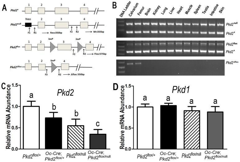
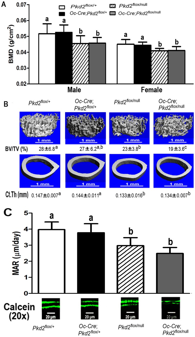
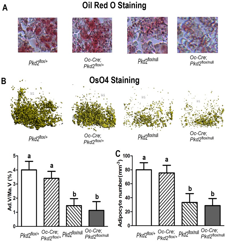
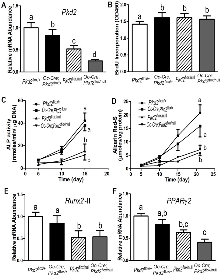
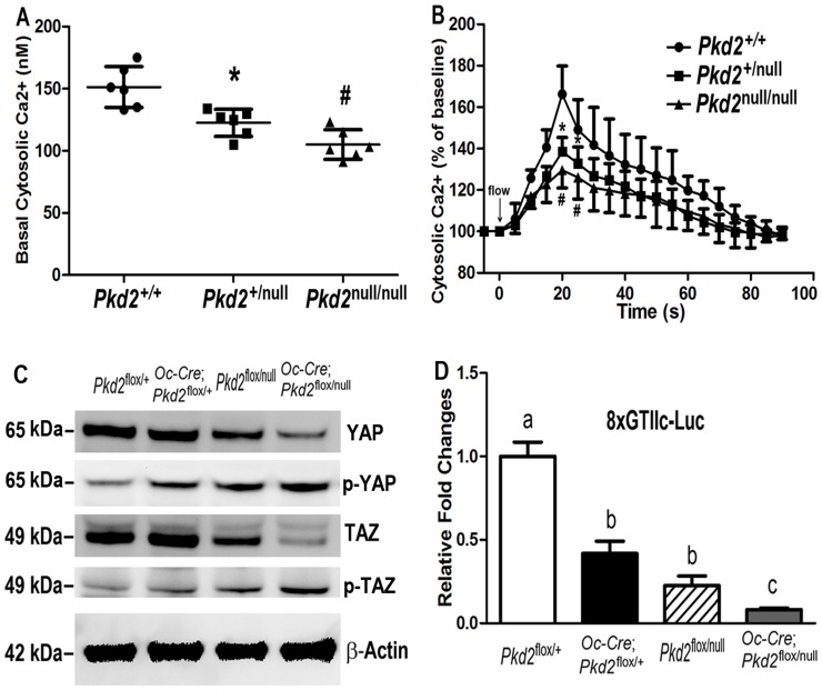
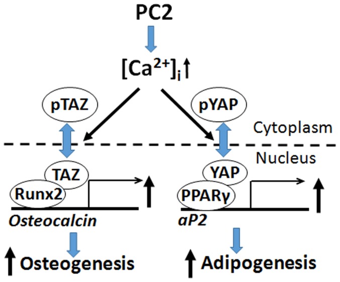
Polycystin-1 (Pkd1) interacts with polycystin-2 (Pkd2) to form an interdependent signaling complex. Selective deletion of Pkd1 in the osteoblast lineage reciprocally regulates osteoblastogenesis and adipogenesis. The role of Pkd2 in skeletal development has not been defined. To this end, we conditionally inactivated Pkd2 in mature osteoblasts by crossing Osteocalcin (Oc)-Cre;Pkd2+/null mice with floxed Pkd2 (Pkd2flox/flox) mice. Oc-Cre;Pkd2flox/null (Pkd2Oc-cKO) mice exhibited decreased bone mineral density, trabecular bone volume, cortical thickness, mineral apposition rate and impaired biomechanical properties of bone. Pkd2 deficiency resulted in diminished Runt-related transcription factor 2 (Runx2) expressions in bone and impaired osteoblastic differentiation ex vivo. Expression of osteoblast-related genes, including, Osteocalcin, Osteopontin, Bone sialoprotein (Bsp), Phosphate-regulating gene with homologies to endopeptidases on the X chromosome (Phex), Dentin matrix protein 1 (Dmp1), Sclerostin (Sost), and Fibroblast growth factor 23 (FGF23) were reduced proportionate to the reduction of Pkd2 gene dose in bone of Oc-Cre;Pkd2flox/+ and Oc-Cre;Pkd2flox/null mice. Loss of Pkd2 also resulted in diminished peroxisome proliferator-activated receptor γ (PPARγ) expression and reduced bone marrow fat in vivo and reduced adipogenesis in osteoblast culture ex vivo. Transcriptional co-activator with PDZ-binding motif (TAZ) and Yes-associated protein (YAP), reciprocally acting as co-activators and co-repressors of Runx2 and PPARγ, were decreased in bone of Oc-Cre;Pkd2flox/null mice. Thus, Pkd1 and Pkd2 have coordinate effects on osteoblast differentiation and opposite effects on adipogenesis, suggesting that Pkd1 and Pkd2 signaling pathways can have independent effects on mesenchymal lineage commitment in bone.


Conflict of interest statement

Competing Interests: The authors have read the journal's policy and the authors of this manuscript have the following competing interests: Dr. Amber Rath Stern is employed by a commercial company, Engineering Systems, Inc. This does not alter the authors' adherence to PLOS ONE policies on sharing data and materials.

Figures

Figure 1
Figure 1. Osteocalcin(Oc)-Cre-mediated bone specific deletion of Pkd2 from the floxed Pkd2 allele (Pkd2 flox).
(A) Schematic illustration of wild-type (Pkd2 +), null (Pkd2 null, deleted Exon 1), and floxed Pkd2 allele before (Pkd2 flox) and after deletion (Pkd2 Δflox) of the lox P cassette containing Exon 3 via Cre-mediated recombination. (B) Genotype PCR analysis of different tissues that were harvested from 6-week-old Oc-Cre;Pkd2 flox/null mice showed bone specific deletion of the Pkd2 gene. Osteocalcin-Cre-mediated recombination of excised floxed Pkd2 (Pkd2 Δflox) allele occurred exclusively in bone, whereas non-skeletal tissues retained the floxed Pkd2 allele (Pkd2 flox). (C and D) Real-time RT-PCR analysis of total Pkd2 and Pkd1 transcripts. Expression of total Pkd2 transcripts was performed using Pkd2- or Pkd1-allele-specific primers as described in Experimental Procedures. The normal Pkd2 + vs cyclophilin A is normalized to the mean ratio of 5 control mice, which has been set to 1. The percentage of conditional and global deleted transcripts was calculated from the relative levels of the normal Pkd2 + transcripts in different Pkd2 exons. Data are mean ±S.D. from 6–8 individual mice. Values sharing the same superscript in C and D are not significantly different at P<0.05.
Figure 2
Figure 2. Pkd2 deficiency results in loss of bone mass.
(A) Effects of Pkd2 deficiency on bone mineral density (BMD) at 6 weeks of age. Compared to control Pkd2 flox/+ mice, conditional heterozygous Oc-Cre;Pkd2 flox/+ mice exhibited no abnormalities in bone mass. However, both global heterozygous Pkd2 flox/null and conditional Pkd2 Oc-cKO null mice were osteopenic, as evidenced by respective 14.4% and 12.5% reduction BMD in male adult mice. (B) Effects of Pkd2 deficiency on structure of femurs. µCT analysis of the distal femoral metaphyses and midshaft diaphyses revealed that the reduction in bone mass in both global heterozygous Pkd2 flox/null and conditional Pkd2 Oc-cKO null mice were caused by reductions in trabecular bone volume (19.3% and 30.9%, respectively) and cortical bone thickness (8.4% and 7.3%, respectively). In contrast, conditional heterozygous Oc-Cre;Pkd2 flox/+ had no significant difference in both trabecular and cortical bone compared with control mice. (C) Effects of Pkd2 deficiency on bone formation rate. Both Pkd2 flox/null and Pkd2 Oc-cKO null mice exhibited a significant decrease in periosteal mineral apposition rate (MAR, 25.1% and 37.3%, respectively), indicating a reduction of bone formation rate in Pkd2 deficient mice. Data represent the mean±S.D. from 6–8 individual mice. Values sharing the same superscript in different group are not significantly different at P<0.05.
Figure 3
Figure 3. Pkd2 deficiency impairs adipogenesis in bone.
(A) Histology of adipocytes in decalcified tibias. Oil Red O staining showed that the numbers of adipocytes and fat droplets in tibial bone marrow were much less in 20-week-old Pkd2 flox/null and Pkd2 Oc-cKO null mice compared with age-matched control Pkd2 flox/+ and Oc-Cre;Pkd2 flox/+ mice. (B) Osmium tetroxide (OsO4) staining of decalcified femurs by µCT analyses. Upper panel showed the representative images of distal femoral bone marrow by OsO4 staining (yellow). Lower panel displayed adipocyte volume/marrow volume (Ad.V/Ma.V, %) and adipocyte number (Ad.N, mm−3) by calculation. Consistent with Oil Red O staining, µCT analyses showed that adipocyte volume/marrow volume (Ad.V/Ma.V, %) and adipocyte number (Ad.N, mm−3) were much lower in the distal femurs from 20-week-old Pkd2 flox/null and Pkd2 Oc-cKO null mice compared with age-matched control Pkd2 flox/+ and Oc-Cre;Pkd2 flox/+ mice, indicating an impairment of adipogenesis in the Pkd2 deficient mice. Data represent the mean±S.D. from 6–8 individual mice. Values sharing the same superscript in different group are not significantly different at P<0.05.
Figure 4
Figure 4. Pkd2 deficiency osteoblasts have a developmental defect in vitro.
(A) A real-time RT-PCR analysis of total Pkd2 transcripts in osteoblast cultures. A gene dose-dependent reduction of Pkd2 transcripts was observed in immortalized control and Pkd2-deficient osteiblasts. (B) BrdU incorporation. Primary cultured Pkd2 deficient osteoblasts exhibited a higher BrdU incorporation than control Pkd2 flox/+ osteoblasts for 6 hours, indicating increased proliferation rate in the Pkd2-deficient osteoblasts. (C) ALP activity. Primary cultured Pkd2 flox/null and Pkd2 Oc-cKO null osteoblasts displayed time-dependent increments in alkaline phosphatase (ALP) activities during 15 days of culture, but the ALP activity was significantly lower at different time points compared with control Pkd2 flox/+ and Oc-Cre;Pkd2 flox/+ osteoblasts. (D) Quantification of mineralization. Alizarin Red-S was extracted with 10% cetylpyridinium chloride and quantified as described in Experimental Procedures. Primary cultured Pkd2 flox/null and Pkd2 Oc-cKO null had time-dependent increments in Alizarin Red-S accumulation during 22 days of culture, but the accumulation was significantly lower at different time points compared with control Pkd2 flox/+ and Oc-Cre;Pkd2 flox/+ osteoblasts. (E and F) Gene expression profiles by real-time RT-PCR. 10-days cultured Pkd2 flox/null and Pkd2 Oc-cKO null osteoblasts in osteogenic differentiation media showed a significant attenuation in both osteogenesis and adipogenesis compared to control Pkd2 flox/+ and Oc-Cre;Pkd2 flox/+, evidenced by a significant reduction in osteoblastic and adipogenic markers, such as Runx2-II and PPARγ2. Data are mean±S.D. from triple three independent experiments. Values sharing the same superscript in different group are not significantly different at P<0.05.
Figure 5
Figure 5. Signaling pathways in Pkd2 deficiency osteoblasts.
(A) Basal intracellular calcium ([Ca2+]i) levels. Heterozygous Pkd2 +/null osteoblasts (n = 6) showed a significantly lower basal [Ca2+]i levels compared with wild-type Pkd2 +/+ osteoblasts (n = 6), and homozygous Pkd2 null/null osteoblasts (n = 6) had greater reductions of basal [Ca2+]i compared with the heterozygous Pkd2 +/null osteoblasts (n = 6). (B) Flow-induced [Ca2+]i response. A gene dose-dependent reduction of flow-induced [Ca2+]i response was observed in the Pkd2-deficient osteoblasts compared with wild-type Pkd2 +/+ osteoblasts, indicating an impairment of calcium channel activity in the Pkd2-deficient osteoblasts. (C) TAZ/YAP-dependent transcriptional activation as assessed by 8xGTIIc-luciferase activity. A gene dose-dependent reduction of basal TAZ/YAP activity was observed the in immortalized primary osteoblasts derived from the Pkd2-deficient osteoblasts compared to the wild-type controls. (D) Western blot analysis. Phosphorylation of YAP was significantly increased, while the amount of TAZ was significantly decreased in Pkd2-deficient osteoblasts than in wild-type controls. Data are mean±S.D. from triple independent experiments. Values sharing the same superscript in different group are not significantly different at P<0.05.
Figure 6
Figure 6. Schema showing potential interactions between polycystins and Hippo signaling pathways in osteoblasts.
PC-2 coordinately regulates PPARγ and Runx2 to respectively control adipogenesis and osteoblastogenesis. Hippo signaling effectors Yap and Taz are also coordinately regulated by PC-2 as well as other physical forces that act as co-factors for PPARγ and Runx2. Inverse effects on osteoblast and adipocyte differentiation by PC-1 might be explained by uncoupling PC-1 and PC-2 signaling leading to enhancement of PC-1 C-terminal tail (PC-1-CTT)/Taz signaling and increased Runx2-dependent osteoblastogenesis and decreased PPARγ-mediated adipogenesis.

References

    1. Qian F, Germino FJ, Cai Y, Zhang X, Somlo S, et al. (1997) PKD1 interacts with PKD2 through a probable coiled-coil domain. Nat Genet 16:179–183. - PubMed
    1. Tsiokas L, Kim E, Arnould T, Sukhatme VP, Walz G (1997) Homo- and heterodimeric interactions between the gene products of PKD1 and PKD2. Proc Natl Acad Sci U S A 94:6965–6970. - PMC - PubMed
    1. Hanaoka K, Qian F, Boletta A, Bhunia AK, Piontek K, et al. (2000) Co-assembly of polycystin-1 and -2 produces unique cation-permeable currents. Nature 408:990–994. - PubMed
    1. Yu Y, Ulbrich MH, Li MH, Buraei Z, Chen XZ, et al. (2009) Structural and molecular basis of the assembly of the TRPP2/PKD1 complex. Proc Natl Acad Sci U S A 106:11558–11563. - PMC - PubMed
    1. Zhu J, Yu Y, Ulbrich MH, Li MH, Isacoff EY, et al. (2011) Structural model of the TRPP2/PKD1 C-terminal coiled-coil complex produced by a combined computational and experimental approach. Proc Natl Acad Sci U S A 108:10133–10138. - PMC - PubMed

Publication types

Substances