. 2014 Dec 2;9(12):e112316.
doi: 10.1371/journal.pone.0112316. eCollection 2014.

Technical complications during veno-venous extracorporeal membrane oxygenation and their relevance predicting a system-exchange--retrospective analysis of 265 cases

Affiliations

Technical complications during veno-venous extracorporeal membrane oxygenation and their relevance predicting a system-exchange--retrospective analysis of 265 cases

Matthias Lubnow et al. PLoS One. .

Abstract

Objectives: Technical complications are a known hazard in veno-venous extracorporeal membrane oxygenation (vvECMO). Identifying these complications and predictive factors indicating a developing system-exchange was the goal of the study.

Methods: Retrospective study on prospectively collected data of technical complications including 265 adult patients (Regensburg ECMO Registry, 2009-2013) with acute respiratory failure treated with vvECMO. Alterations in blood flow resistance, gas transfer capability, hemolysis, coagulation and hemostasis parameters were evaluated in conjunction with a system-exchange in all patients with at least one exchange (n = 83).

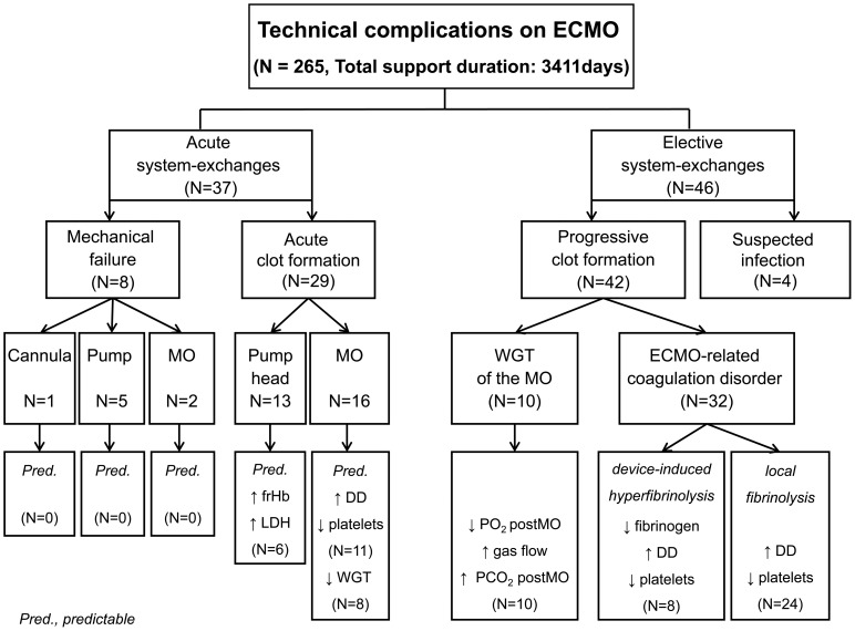
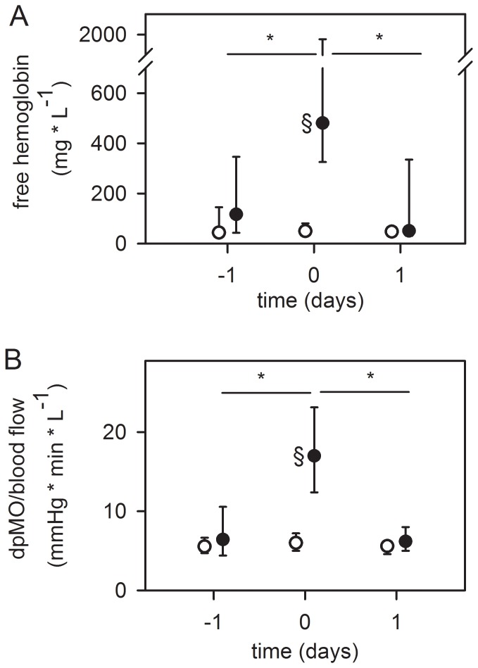
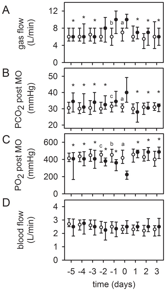
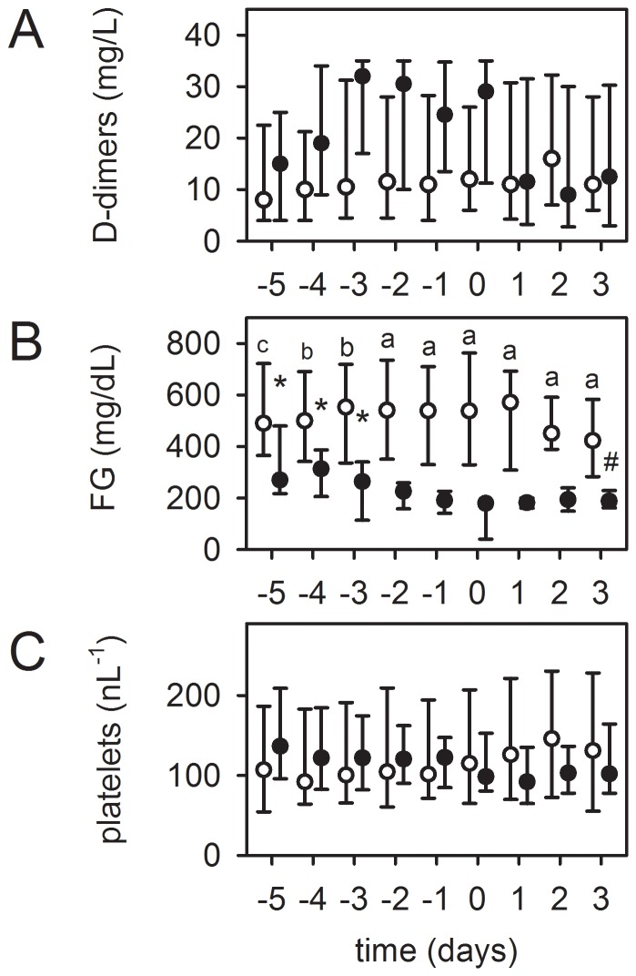
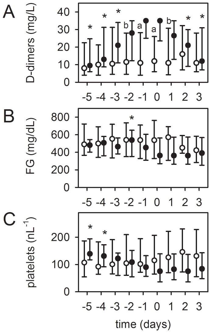
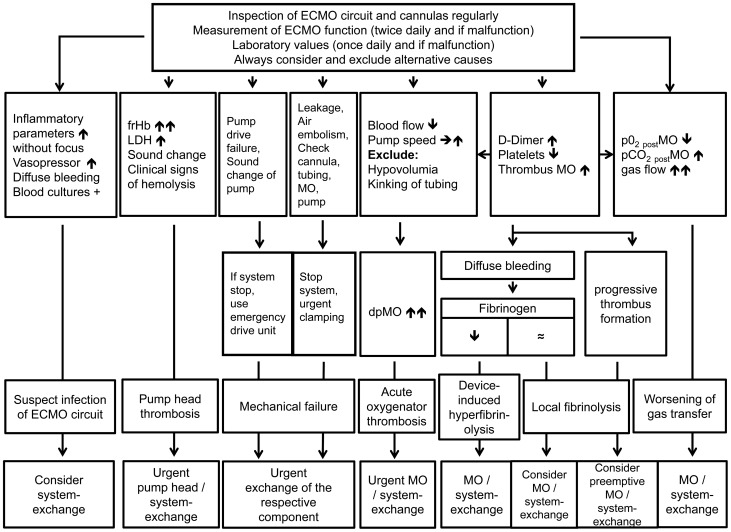
Results: Values presented as median (interquartile range). Patient age was 50(36-60) years, the SOFA score 11(8-14.3) and the Murray lung injury Score 3.33(3.3-3.7). Cumulative ECMO support time 3411 days, 9(6-15) days per patient. Mechanical failure of the blood pump (n = 5), MO (n = 2) or cannula (n = 1) accounted for 10% of the exchanges. Acute clot formation within the pump head (visible clots, increase in plasma free hemoglobin (frHb), serum lactate dehydrogenase (LDH), n = 13) and MO (increase in pressure drop across the MO, n = 16) required an urgent system-exchange, of which nearly 50% could be foreseen by measuring the parameters mentioned below. Reasons for an elective system-exchange were worsening of gas transfer capability (n = 10) and device-related coagulation disorders (n = 32), either local fibrinolysis in the MO due to clot formation (increased D-dimers [DD]), decreased platelet count; n = 24), or device-induced hyperfibrinolysis (increased DD, decreased fibrinogen [FG], decreased platelet count, diffuse bleeding tendency; n = 8), which could be reversed after system-exchange. Four MOs were exchanged due to suspicion of infection.

Conclusions: The majority of ECMO system-exchanges could be predicted by regular inspection of the complete ECMO circuit, evaluation of gas exchange, pressure drop across the MO and laboratory parameters (DD, FG, platelets, LDH, frHb). These parameters should be monitored in the daily routine to reduce the risk of unexpected ECMO failure.


Conflict of interest statement

Competing Interests: CS is an advisor for Maquet Cardiopulmonary AG, Rastatt, Germany. ML, AP and TM received travel support and lecture honoraries from Maquet Cardiopulmonary AG, Rastatt, Germany. TB is member of the advisory board of Novalung, Heilbronn, Germany and he received honoraria. None of the other authors has a financial relationship with a commercial entity that has an interest in the subject of the presented manuscript or other conflicts of interest to disclose. This does not alter the authors' adherence to PLOS ONE policies on sharing data and materials.

Figures

Figure 1
Figure 1. Retrospective analysis of reasons for acute and elective system-exchange during vvECMO.
Acute exchanges were all events that required an immediate intervention of perfusionists. Mechanical failure was defined as leakage at the MO, pump head problems, dysfunction of the pumpdrive, cannula or circuit rupture. Acute clot formation within the MO caused a severe increase in the pressure drop across the MO (dpMO) followed by a decrease in blood flow, while acute clot formation in the pump head was indicated by a dramatic increase in plasma free hemoglobin concentration. Suspected infection based on clinical observations (see text). Progressive clot formation was observed in almost all remaining MOs, which caused a worsened gas exchange capability of the MO (decrease in pO2 postMO, increase in pCO2 postMO, increase in sweep gas flow) and an alteration in coagulation parameters (increase in D-dimer levels, decrease in fibrinogen levels and decrease in platelet counts).
Figure 2
Figure 2. Pump head thrombosis (top, A) and acute oxygenator thrombosis (bottom, B).
(A) Alterations in plasma free hemoglobin concentration in patients with pump head thrombosis one day before, at system-exchange and one day thereafter (n = 13, black dots). (B) Alterations in pressure drop across the oxygenator (dpMO) normalized by blood flow in patients with acute oxygenator thrombosis one day before, at system-exchange and one day thereafter (n = 16, black dots). Circles in both graphs: Patients without system exchange and a comparable support time of the MO (≥12 days, n = 36) were used as control group. Since the system-exchange was necessary after 9 (6–12) days (Table 2), values at day 9 after starting ECMO therapy were set as “day 0” and days −1 and 1 depicted accordingly. Data are presented as median and interquartile range. *, p≤0.05 compared to day 0 (refers to black dots); §, p≤0.001 compared to patients without system exchange (circles).
Figure 3
Figure 3. Worsened gas exchange capability as a reason for an elective system-exchange.
Timeline of respective values before and after sytem-exchange, “day 0” =  system-exchange. Patients with system-exchange (black dots, n = 10). Despite an increase in gas flow rate (100% O2) (top, A), the partial pressure of CO2 at the outlet of the MO (pCO2 postMO) increased significantly (>40 mmHg) (middle, B). The oxygenation capability (pO2 postMO) decreased (middle, C). ECMO blood flow remained unchanged (bottom, D). After system-exchange, gas exchange data improved significantly. Circles in all graphs: Patients without system-exchange and a comparable support time of the MO (≥12 days, n = 36) were used as control group. Since the system-exchange was necessary after 9 (6–12) days (Table 2), values at day 9 after starting ECMO therapy were set as “day 0” and depicted accordingly. Data are presented as median and interquartile range. *, p≤0.05 compared to day 0 (refers to black dots). a, p≤0.001; b, p≤0.01; c, p≤0.05 compared to patients without system-exchange (circles).
Figure 4
Figure 4. Device-induced hyperfibrinolysis as a reason for an elective system-exchange.
Timeline of respective values before and after sytem-exchange, “day 0” =  system-exchange. Patients with system-exchange (black dots, n = 8): (B) Fibrinogen (FG) concentration decreased significantly below the normal value of 200 mg/dl, while (A) levels of D-dimers visually increased and (C) platelet count decreased (both not statistically significant). After system-exchange the coagulation disorder could be stopped. Circles: Patients without exchange and comparable support time (≥12 days, n = 36). Data at day 9 after starting ECMO therapy were set as “day 0”, other values depicted accordingly. Data are presented as median and interquartile range. *, p≤0.05 compared to day 0 (refers to black dots). Differences between patients with and without exchange on respective days (a, p≤0.001; b, p≤0.01; c, p≤0.05).
Figure 5
Figure 5. Clot formation and local fibrinolysis in the MO as a reason for an elective system-exchange.
Timeline of respective values before and after sytem-exchange, “day 0”  =  system-exchange. In 24 patients (black dots), the MO was exchanged after a significant increase in D-dimer levels (A) without relevant alteration of fibrinogen (FG) levels (B). At the same time, platelet count decreased (C). After exchange, D-dimer concentration decreased and platelet counts remained stable. Circles: Patient population without exchange and comparable support time (≥12 days, n = 36). Data at day 9 after starting ECMO therapy were set as “day 0”, other values depicted accordingly. Data are presented as median and interquartile range. *, p≤0.05 compared to day 0 (refers to black dots). Differences between patients with and without exchange on respective days (a, p≤0.001; b, p≤0.01).
Figure 6
Figure 6. Flowchart for detecting and managing technical complications on ECMO.
Not all predictors have to be present. Differential diagnoses always have to be ruled out before pathologic values can be attributed to a failing ECMO system. Different types of failure can appear together and the decision for making an exchange is often multifactorial. Therefore, usually irrespective of the leading cause of technical failure, a complete system-exchange is done, as seen in this study. If respective components are available and only one single technical failure is present, it is also possible to exchange only the failing component.

References

    1. MacLaren G, Combes A, Bartlett RH (2012) Contemporary extracorporeal membrane oxygenation for adult respiratory failure: life support in the new era. Intensive Care Med 38:210–220. - PubMed
    1. Peek GJ, Elbourne D, Mugford M, Tiruvoipati R, Wilson A, et al. (2010) Randomised controlled trial and parallel economic evaluation of conventional ventilatory support versus extracorporeal membrane oxygenation for severe adult respiratory failure (CESAR). Health Technol Assess 14:1–46. - PubMed
    1. Peek GJ, Mugford M, Tiruvoipati R, Wilson A, Allen E, et al. (2009) Efficacy and economic assessment of conventional ventilatory support versus extracorporeal membrane oxygenation for severe adult respiratory failure (CESAR): a multicentre randomised controlled trial. Lancet 374:1351–1363. - PubMed
    1. Australia, New Zealand Extracorporeal Membrane Oxygenation Influenza I Davies A, Jones D, Bailey M, et al. (2009) Extracorporeal Membrane Oxygenation for 2009 Influenza A(H1N1) Acute Respiratory Distress Syndrome. JAMA 302:1888–1895. - PubMed
    1. Combes A, Pellegrino V (2011) Extracorporeal membrane oxygenation for 2009 influenza A (H1N1)-associated acute respiratory distress syndrome. Semin Respir Crit Care Med 32:188–194. - PubMed

Publication types