. 2015 Mar;66(5):1245-57.
doi: 10.1093/jxb/eru475. Epub 2014 Dec 1.

Arabinogalactan proteins are involved in root hair development in barley

Affiliations

Arabinogalactan proteins are involved in root hair development in barley

Marek Marzec et al. J Exp Bot. 2015 Mar.

Abstract

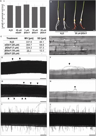
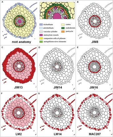
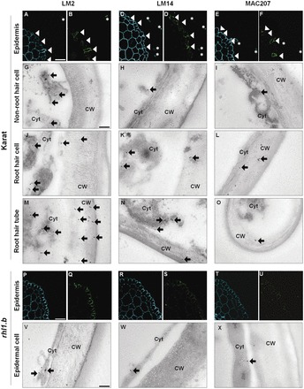
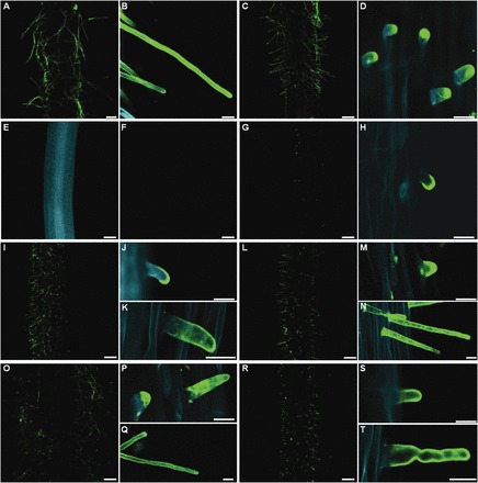
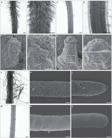
The arabinogalactan proteins (AGPs) are involved in a range of plant processes, including cell differentiation and expansion. Here, barley root hair mutants and their wild-type parent cultivars were used, as a model system, to reveal the role of AGPs in root hair development. The treatment of roots with different concentrations of βGlcY (a reagent which binds to all classes of AGPs) inhibited or totally suppressed the development of root hairs in all of the cultivars. Three groups of AGP (recognized by the monoclonal antibodies LM2, LM14, and MAC207) were diversely localized in trichoblasts and atrichoblasts of root hair-producing plants. The relevant epitopes were present in wild-type trichoblast cell walls and cytoplasm, whereas in wild-type atrichoblasts and in all epidermal cells of a root hairless mutant, they were only present in the cytoplasm. In all of cultivars the higher expression of LM2, LM14, and MAC207 was observed in trichoblasts at an early stage of development. Additionally, the LM2 epitope was detected on the surface of primordia and root hair tubes in plants able to generate root hairs. The major conclusion was that the AGPs recognized by LM2, LM14, and MAC207 are involved in the differentiation of barley root epidermal cells, thereby implying a requirement for these AGPs for root hair development in barley.

Keywords: Arabinogalactan proteins (AGPs); Yariv.; barley (Hordeum vulgare); cell differentiation; monoclonal antibodies; root hairs.


Figures

Fig. 1.
Fig. 1.
The effect of βGlcY treatment on root growth and root hair differentiation in barley cv. Karat. (A, B) Root length was not significantly influenced, while (C–J) root hair development was inhibited. (C) Root hair lengths estimated from at least 1000 root hairs sampled from 15 roots. (D) The response of root epidermal cells to 25 µM βGlcY. (E–J) Light microscopy analysis: (E,F) hairless mutant roots exposed to 25 µM βGlcY; (G–H) primordia only producing mutant roots exposed to 10 µM βGlcY; (I) treatment with 1 µM βGlcY had no effect on root hair elongation; (J) a control treatment with 25 µM αGalY. Asterisks, shorter epidermal cells; arrows, bulging cells; arrowheads, primordia; MV, mean value; SD, standard deviation. Scale bars in (B) 1cm, (D) 100 µm, (E, G, and I–J) 200 µm; (F, H) 20 µm. Underlined mean value in table indicate the statistical significance, in comparison to control conditions (Student’s t test (P < 0.05)).
Fig. 2.
Fig. 2.
Schematic overview of AGP epitope distribution derived from transverse sections made from the mature root hair zone. (A) The organization of the root. (B–H) Abundance of the various AGP epitopes. (B) JIM8 in the endodermis and metaphloem sieve elements. (C) JIM13 was distributed throughout the root except in the metaxylem. (D) JIM14 was restricted to the metaphloem sieve elements, whereas (E) JIM16 was restricted to the endodermis. The three epitopes (F) LM2, (G) LM14, and (H) MAC207 were heterogeneously distributed in the epidermis. Signal strength indicated by colour: dark red (strong), light red (weak), and white (none).
Fig. 3.
Fig. 3.
Immunolocalization of LM2, LM14, and MAC207 epitopes in the rhizodermis of barley cv. Karat and the rhl1.b mutant. (A, C, E, P, R, T) Autofluorescence illustrates cell patterning in the mature root hair zone. Fluorescence labelling of AGP epitopes in (B, D, F) cv. Karat and (Q, S, U) the rhl1.b mutant, with (B, D, F, Q, S, U, G–O, V–X) showing subcellular localization based on immunogold labelling. (A–F) Epitopes were more abundant in the trichoblasts and root hair tubes than in the atrichoblasts. (G–O) In the trichoblast cell wall, LM2, LM14, and MAC207 epitopes were only detected in the wild-type cultivars (H–X). In the root hairless mutant, the three epitopes were homogeneously distributed within the epidermis. Asterisks, root hair tubes; arrowheads, trichoblasts; arrows, gold particles; CW, cell wall; Cyt, cytoplasm. Scale bars: (A–F and P–U) 50 µm; (G–O and V–X), 100nm.
Fig. 4.
Fig. 4.
The localization of LM2 epitopes in whole-mount immunolabelled root sections of barley cultivars Karat and Dema, and the root hair mutants rhl1.b, rhp1.a, rhs1.a, 2.a, 3.a, and 4.a. Epitope was detected (A, B) on the surface of cv. Karat root-hair tubes and (C, D) in the zone harbouring root hairs in cv. Dema. (E) Autofluorescence in rhl1.b and (F) the lack of any epitope on the root surface. (G, H) Clear signal in the primordia formed by rhp1.a. (I) Epitope in rhs1.a, focusing on (J) young and (K) mature root hairs. Similar comparisons are shown for (L–N) rhs2.a, (O–Q) rhs3.a, (R–T) rhs4.a. Scale bar in (A, C, E–G, I, L, O, R) 200 µm, and in (B, D, H, J, K, M, N, P, Q, S, T) 20 µm.
Fig. 5.
Fig. 5.
LM2 epitope distribution on the barley cv. Karat root hair surface as shown by immunogold labelling visualized by (A–D, J, M) light microscopy and (C–I, K, L, N, O) SEM. (A, B) Negative control (no primary antibody); (C, D) LM2 signal following the inclusion of gold-conjugated secondary antibody; (E) primordium; (F) detail of the primordium tip. Label strength decreased from (H) the tip to (I) the base of a growing root hair (G). (J) Root hairs displaying an even distribution of LM2 epitopes in (K) the central part and (L) the tip of fully developed root hairs. (M–O) No epitopes were detected in the negative control. Scale bar in (A–D, J, M) 200 µm, and in (E–L, N, O) 2 µm.

References

    1. Andeme-Onzighi C, Sivaguru M, Judy-March J, Baskin TI, Driouich A. 2002. The reb1-1 mutation of Arabidopsis alters the morphology of trichoblasts, the expression of arabinogalactan proteins and the organisation of cortical microtubules. Planta 215, 949–958. - PubMed
    1. Bradley DJ, Wood EA, Larkins AP, Galfre G, Butcher GW, Brewin NJ. 1988. Isolation of monoclonal antibodies reacting with peribacteroid membranes and other components of pea root nodules containing Rhizobium leguminosarum . Planta 173, 149–160. - PubMed
    1. Casero PJ, Casimiro I, Knox JP. 1998. Occurrence of cell surface arabinogalactan-protein and extensin epitopes in relation to pericycle and vascular tissue development in the root apex of four species. Planta 204, 252–259.
    1. Chen T, Teng N, Wu X, Wang Y, Tang W, Samaj J, Baluska F, Lin J. 2007. Disruption of actin filaments by latrunculin B affects cell wall construction in Picea meyeri pollen tube by disturbing vesicle trafficking. Plant and Cell Physiology 1, 19–30. - PubMed
    1. Chmielewska B, Janiak A, Karcz J, et al. 2014. Morphological, genetic and molecular characteristics of barley root hair mutants. Journal of Applied Genetics 10.1007/s13353-014-0225-x. - PMC - PubMed

Publication types

LinkOut - more resources