Review
. 2015 Jan;16(1):57-64.
doi: 10.1038/nrm3916. Epub 2014 Dec 3.

Regulation of kinetochore-microtubule attachments through homeostatic control during mitosis

Affiliations
Review

Regulation of kinetochore-microtubule attachments through homeostatic control during mitosis

Kristina M Godek et al. Nat Rev Mol Cell Biol. 2015 Jan.

Abstract

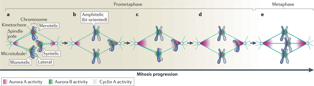
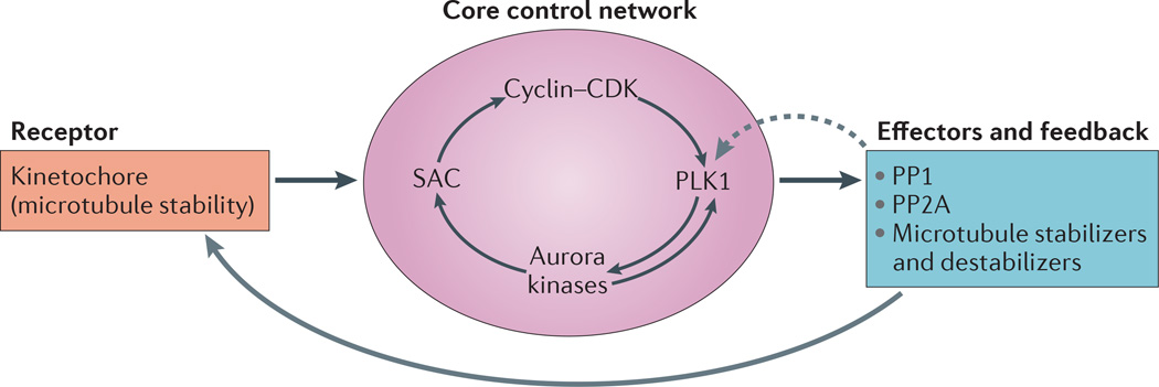
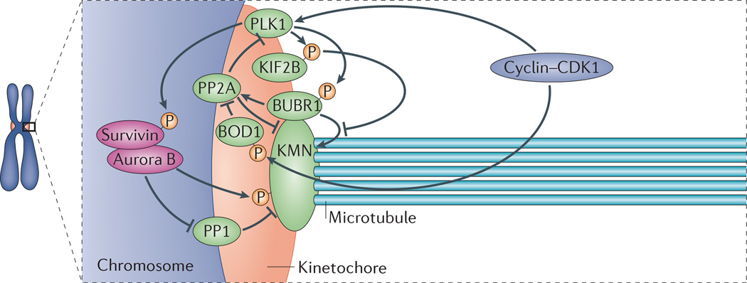
Faithful chromosome segregation during mitosis is essential for genome integrity and is mediated by the bi-oriented attachment of replicated chromosomes to spindle microtubules through kinetochores. Errors in kinetochore-microtubule (k-MT) attachment that could cause chromosome mis-segregation are frequent and are corrected by the dynamic turnover of k-MT attachments. Thus, regulating the rate of spindle microtubule attachment and detachment to kinetochores is crucial for mitotic fidelity and is frequently disrupted in cancer cells displaying chromosomal instability. A model based on homeostatic principles involving receptors, a core control network, effectors and feedback control may explain the precise regulation of k-MT attachment stability during mitotic progression to ensure error-free mitosis.


Figures

Figure 1
Figure 1. Kinetochore–microtubule attachments in mitosis
Different types of kinetochore–microtubule (k–MT) attachments occur in prometaphase (a–d). These include transient intermediates such as monotelic attachments (in which only one of the sister kinetochores is attached to microtubules from one spindle pole) and lateral attachments (in which kinetochores are bound to the side wall of microtubules). In addition, errors in attachment exist, including syntelic attachments (in which both sister kinetochores are attached to microtubules from the same spindle pole) and merotelic attachments (in which a single kinetochore is attached to microtubules from both spindle poles). As cells progress through mitosis, the erroneous attachments are corrected, leading to end-on, bi-oriented attachments, in which sister kinetochores are attached to microtubules from opposite spindle poles to support faithful chromosome segregation. A core control network regulates the stability of k–MT attachments to promote efficient error correction and ensure faithful chromosome segregation. Note that, for simplicity, only cyclin A, Aurora A kinase and Aurora B kinase — which are key components of the core control network — are shown in the figure. Cyclin A forms a temporal gradient as its abundance declines during prometaphase (a–d), whereas Aurora A and Aurora B kinases form spatial gradients at spindle poles and at centromeres, respectively. Correction of syntely involves the recognition and targeted destabilization of k–MT attachments through the combined activities of Aurora A and Aurora B kinases as chromosomes are pulled towards the spindle poles (a-d). The release of microtubules from kinetochores permits the chromosome to move to the spindle midzone through lateral k–MT attachments to re-establish bi-oriented attachments (c,d). Correction of merotely involves the indiscriminate destabilization of k–MT attachments on aligned chromosomes during prometaphase (a to b, c to d). The high detachment rate of k–MT attachments in prometaphase (dashed lines) that is ensured by cyclin A activity combines with the back-to-back geometry of sister kinetochores to facilitate bi-oriented attachments. In metaphase (e), k–MT attachments switch to more stable attachments (solid lines) as cyclin A levels fall below a critical threshold.
Figure 2
Figure 2. Homeostatic control circuit for regulating kinetochore–microtubule attachment stability in mitosis
A receptor, a core control network, effector modules and feedback regulatory mechanisms (grey arrows) of a homeostatic control system regulate kinetochore–microtubule (k–MT) attachment stability. Conserved kinetochore proteins directly bind microtubules, thus forming the receptor. These probably include the proteins of the KMN network (BOX 1). Kinetochore proteins respond to microtubule attachment stability to send signals to the interactive core control network, which is composed of the spindle assembly checkpoint (SAC) and cyclin–cyclin-dependent kinase (CDK) complexes, polo-like kinase 1 (PLK1) and Aurora kinases (and possibly others, including the inner centromere protein shugoshin (SGO1) and haspin kinase). Arrows inside the core control network reflect some of the known functional interactions among these components. This network integrates input from the cell cycle regulatory machinery and the microtubule occupancy status of kinetochores to regulate the activity of effector molecules to increase or decrease the stability of k–MT attachments accordingly (solid grey arrow). The system is regulated by negative feedback from protein phosphatases PP1 and PP2A (dashed grey arrow) to enable control in the context of changing environmental conditions; that is, mitotic phase transitions.
Figure 3
Figure 3. The network of regulatory components at kinetochores
The complexity of the signalling pathways acting to regulate kinetochore–microtubule (k–MT) attachment stability is displayed in a nonspecific stage of mitosis (not necessarily prometaphase or metaphase). The KMN network (BOX 1) provides the primary microtubule-binding element in the kinetochore. The core control network — which is composed of polo-like kinase 1 (PLK1), Aurora B kinase and cyclin–cyclin-dependent kinases (CDKs) (and possibly other components, including the inner centromere protein shugoshin (SGO1) and haspin kinase) — acts to regulate the stability of k–MT attachments. The phosphorylation status of their substrates (such as kinesin family member 2B (KIF2B), BUB1-related kinase 1 (BUBR1), bi-orientation of chromosomes in cell division 1 (BOD1) and survivin) is determined by feedback from the phosphatases PP1 and PP2A. The combined activities of the core control proteins and phosphatases determines the relative activities of effector proteins such as the k–MT stabilizer BUBR1 and the k–MT destabilizer KIF2B, which collectively modulate k–MT attachment stability.

References

    1. Cimini D, Degrassi F. Aneuploidy: a matter of bad connections. Trends Cell Biol. 2001;15:442–451. - PubMed
    1. Cimini D. Merotelic kinetochore orientation, aneuploidy, and cancer. Biochim. Biophys. Acta. 2008;1786:32–40. - PubMed
    1. Weaver BA, Cleveland DW. Aneuploidy: instigator and inhibitor of tumorigenesis. Cancer Res. 2006;67:10103–10105. - PMC - PubMed
    1. Siegel JJ, Amon A. New insights into the troubles of aneuploidy. Annu. Rev. Cell Devel. Biol. 2012;28:189–214. - PMC - PubMed
    1. Duijf PH, Benezra R. The cancer biology of whole-chromosome instability. Oncogene. 2013;32:4727–4736. - PubMed

Publication types