

Circulating microparticles in patients with antiphospholipid antibodies: characterization and associations
- PMID: 25467081
- PMCID: PMC4272916
- DOI: 10.1016/j.thromres.2014.11.011
Circulating microparticles in patients with antiphospholipid antibodies: characterization and associations
Abstract
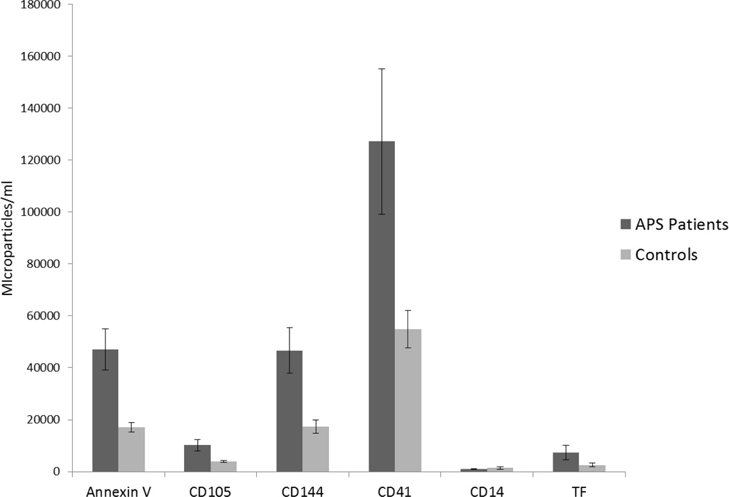
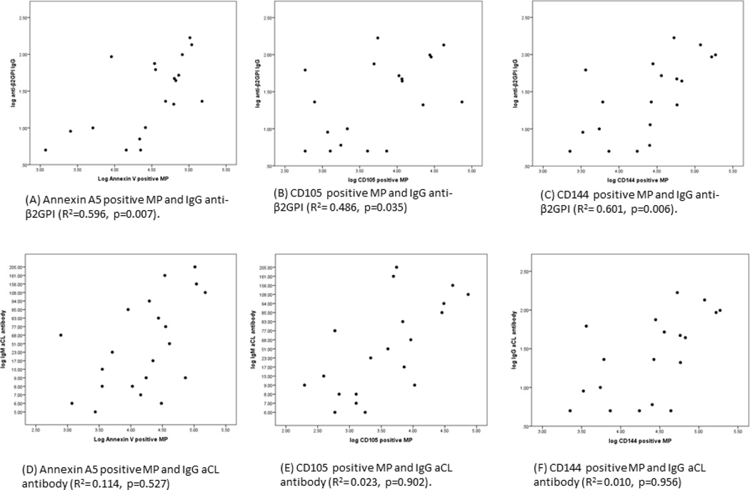
The antiphospholipid syndrome is characterized by venous or arterial thrombosis and/or recurrent fetal loss in the presence of circulating antiphospholipid antibodies. These antibodies cause activation of endothelial and other cell types leading to the release of microparticles with procoagulant and pro-inflammatory properties. The aims of this study were to characterize the levels of endothelial cell, monocyte or platelet derived, and tissue factor-bearing microparticles in patients with antiphospholipid antibodies, to determine the association of circulating microparticles with anticardiolipin and anti-β2-glycoprotein antibodies, and to define the cellular origin of microparticles that express tissue factor. Microparticle content within citrated blood from 47 patients with antiphospholipid antibodies and 144 healthy controls was analyzed within 2hours of venipuncture. Levels of Annexin-V, CD105 and CD144 (endothelial derived), CD41 (platelet derived) and tissue factor positive microparticles were significantly higher in patients than controls. Though levels of CD14 (monocyte-derived) microparticles in patient plasma were not significantly increased, increased levels of CD14 and tissue factor positive microparticles were observed in patients. Levels of microparticles that stained for CD105 and CD144 showed a positive correlation with IgG (R=0.60, p=0.006) and IgM anti-beta2-glycoprotein I antibodies (R=0.58, p=0.006). The elevation of endothelial and platelet derived microparticles in patients with antiphospholipid antibodies and their correlation with anti-β2-glycoprotein I antibodies suggests a chronic state of vascular cell activation in these individuals and an important role for β2-glycoprotein I in development of the pro-thrombotic state associated with antiphospholipid antibodies.
Keywords: Antiphospholipid; Endothelial cell; Microparticles; Platelet; Thrombosis.
Copyright © 2014 Elsevier Ltd. All rights reserved.
Figures

References
-
- Giannakopoulos B, Passam F, Rahgozar S, Krilis SA. Current concepts on the pathogenesis of the antiphospholipid syndrome. Blood. 2007;109:422–429. - PubMed
-
- Rand JH. The antiphospholipid syndrome. Ann Rev Med. 2003;54:409–424. - PubMed
-
- Thiagarajan P, Shapiro SS. Lupus anticoagulants and antiphospholipid antibodies. Hematol Oncol Clin N A. 1998;12:1167–1192. - PubMed
-
- Pierangeli SS, Chen PP, Raschi E, Scurati S, Grossi C, Borghi MO, Palomo I, Harris EN, Meroni PL. Antiphospholipid antibodies and the antiphospholipid syndrome: pathogenic mechanisms. Semin Thromb Hemost. 2008;34:236–250. - PubMed
-
- Ruiz-Irastorza G, Crowther M, Branch W, Khamashta MA. Antiphospholipid syndrome. Lancet. 2010;376:1498–1509. - PubMed
Publication types
MeSH terms
Substances
Grants and funding
LinkOut - more resources
Full Text Sources
Other Literature Sources
Medical
Research Materials
