. 2014 Dec 4;159(6):1417-1432.
doi: 10.1016/j.cell.2014.11.003. Epub 2014 Nov 20.

Identification of spinal circuits transmitting and gating mechanical pain

Affiliations

Identification of spinal circuits transmitting and gating mechanical pain

Bo Duan et al. Cell. .

Abstract

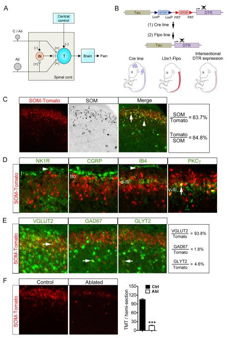
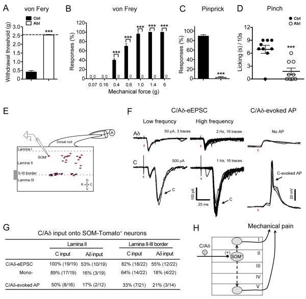
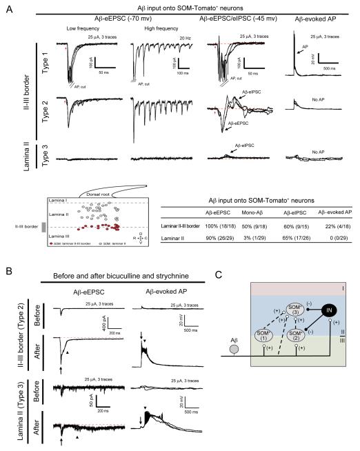
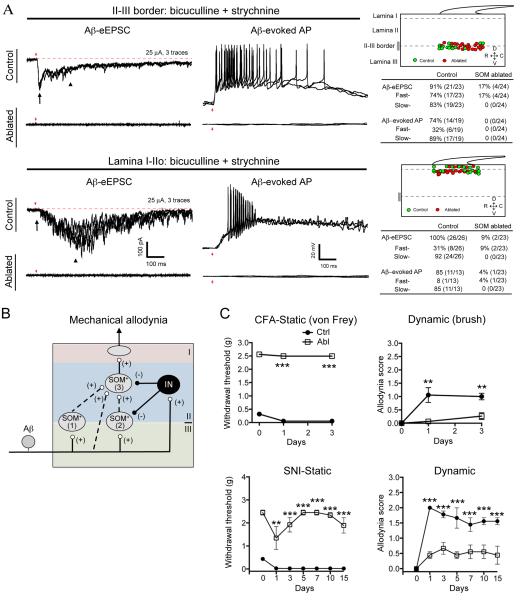
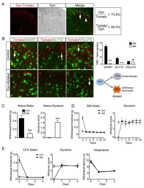
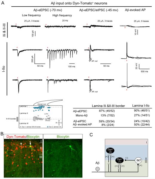
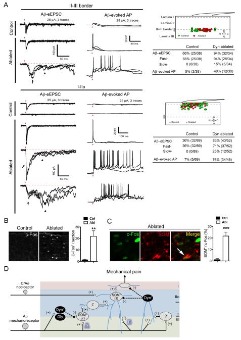
Pain information processing in the spinal cord has been postulated to rely on nociceptive transmission (T) neurons receiving inputs from nociceptors and Aβ mechanoreceptors, with Aβ inputs gated through feed-forward activation of spinal inhibitory neurons (INs). Here, we used intersectional genetic manipulations to identify these critical components of pain transduction. Marking and ablating six populations of spinal excitatory and inhibitory neurons, coupled with behavioral and electrophysiological analysis, showed that excitatory neurons expressing somatostatin (SOM) include T-type cells, whose ablation causes loss of mechanical pain. Inhibitory neurons marked by the expression of dynorphin (Dyn) represent INs, which are necessary to gate Aβ fibers from activating SOM(+) neurons to evoke pain. Therefore, peripheral mechanical nociceptors and Aβ mechanoreceptors, together with spinal SOM(+) excitatory and Dyn(+) inhibitory neurons, form a microcircuit that transmits and gates mechanical pain. PAPERCLIP:


Figures

Figure 1
Figure 1. Intersectional Ablation of SOM lineage Neurons in Spinal Dorsal Horn
(A) Schematics showing the modified gate control theory of pain. “T” represents a spinal pain transmission neuron. “IN”: an inhibitory neuron. “(+)” and “(−)” represent excitatory and inhibitory inputs, respectively. The dashed line from C/Aδ to IN indicates that C/Aδ fibers might activate an unknown pathway to silence IN activity, but this pathway and descending modulation from brain were not studied here. (B) Schematics showing strategy of intersectional ablation in the dorsal spinal cord. “DTR”: diphtheria toxin receptor. (C, D) Double staining of Tomato with SOM mRNA (C) or with other markers (D), on saggital (NK1R) or transverse (others) lumbar spinal sections of adult SOM-Tomato mice. Arrows indicate co-localization. Arrowheads indicate lamina I neurons with singular expression of NK1R or Tomato. (E) Double labeling of Tomato and with indicated mRNAs. Arrows indicate co-localization. (F) Ablation of SOM-Tomato+ neurons in lumbar dorsal spinal cord [105 ± 4 in control (“Ctrl”) group vs 17 ± 2 in ablated (“Abl”) group, n = 15-17 hemi-sections from 3 mice per group; p < 0.001, Student’s unpaired t test]. Data are represented as mean ± SEM. See also Figure S1.
Figure 2
Figure 2. Loss of Acute Mechanical Pain in SOM Abl Mice and C/Aδ Inputs onto SOM-Tomato+ Neurons
(A) Increase of withdrawal thresholds to von Frey fiber stimulation in SOM Abl mice (“Abl”) by up-down method [n = 13 in control (“Ctrl”) group, n = 11 in Abl group; ***, p < 0.001, Student’s unpaired t test]. (B) Reduced withdrawal percentages in SOM Abl mice in response to von Frey filaments (n = 5 in each group; ***, p < 0.001, Student’s unpaired t test) (C) Lost response to pinprick stimulation in SOM Abl mice (n = 13 in Ctrl group, n = 11 in Abl group; ***, p < 0.001, Student’s unpaired t test). (D) Greatly attenuated licking/flinching response to pinching in Abl mice (n = 9 in Ctrl group, n = 9 in Abl group; ***, p < 0.001, Student’s unpaired t test). (E) Schematics showing relative positions of recorded SOM-Tomato+ neurons. (F) Typical traces of C/Aδ-evoked EPSCs and APs showing C/Aδ-fiber inputs onto SOM-Tomato+ neurons. Red arrows indicate stimulation artifacts. (G) The table is a summary of inputs in 41 recorded SOM-Tomato+ neurons from 8 mice. (H) Schematics showing that SOM-Tomato+ neurons in lamina II receive mono-C/Aδ input and transmit noxious signaling to lamina I and/or V pain output neurons, either directly or indirectly (dashed arrows). Data are represented as mean ± SEM. See also Figure S1-S5.
Figure 3
Figure 3. Aβ Input onto SOM-Tomato+ Neurons in Different Spinal Laminae
(A) Aβ input onto SOM-Tomato+ neurons. Upper lane shows three typical traces. Middle showing the relative positions and summary of Aβ inputs onto 47 SOM-Tomato+ neurons from 9 mice. SOM+ neurons are divided into three types (1-3). Red arrows indicate stimulation artifacts. (B) Aβ-evoked EPSCs and EPSPs in type 2 or 3 SOM-Tomato+ neurons before and after bath application of bicuculline (10 μM) and strychnine (2 μM). Arrows and arrowheads indicate fast and slow eEPSCs/eEPSPs/APs, respectively. 4 mice were used. (C) Schematics showing Aβ inputs into types 1-3 of SOM-Tomato+ neurons. “IN”: inhibitory neurons. See also Figure S1.
Figure 4
Figure 4. Loss of Aβ Inputs onto Lamina I/II Neurons and Mechanical Allodynia in SOM Abl mice
(A) Aβ-evoked EPSCs/APs in spinal neurons from control and SOM Abl mice. Left panel shows typical traces and right panel includes the positions of recorded neurons and summary. Red arrows indicate stimulation artifacts. Black arrows and Arrowheads indicate fast and slow eEPSCs, respectively. (B) Schematics showing SOM neurons linking Aβ fibers to lamina I neurons, which is gated by inhibitory neurons. (C) Loss of static (von Frey assay) and dynamic (brush assay) mechanical allodynia following peripheral inflammation and nerve injury in SOM Abl mice (“Abl”, open rectangle) in comparison with control (“Ctrl”, solid circles) (n = 6-7 in each group; p < 0.001, one-way ANOVA with Newman-Keuls post-hoc analysis). Data are represented as mean ± SEM. See also Figure S2.
Figure 5
Figure 5. Spontaneous Development of Mechanical Allodynia in Dyn Abl mice
(A) Double staining of Tomato and Dyn mRNA in the spinal cord of Dyn-Tomato mice. (B) Double staining of Tomato and indicated mRNAs in Dyn-Tomato control mice and Dyn Abl mice. Right panel (upper) is quantification analysis. Schematics in lower right showing selective ablation of inhibitory Dyn lineage neurons. (C) Reduction of withdrawal threshold to static stimuli (von Frey assay) and increase in dynamic allodynia score (brush assay) in Dyn neuron-ablated (“Abl”) mice [n = 15 in control (“Ctrl”) group, n = 10 in Abl group; ***, p < 0.001, Student’s unpaired t test]. (D, E) After spared nerve injury (SNI) (D) or peripheral inflammation by CFA treatment (E), control mice show a reduction in withdrawal thresholds to static stimuli by von Frey assay and an increase of dynamic allodynia score by the brush assay (n = 7, p < 0.001, one-way ANOVA with Newman-Keuls post-hoc analysis). No difference before and after nerve injury or inflammation in Abl mice (n = 6; p > 0.05, one-way ANOVA with Newman-Keuls post-hoc analysis). Data are represented as mean ± SEM. See also Figure S6 and S7.
Figure 6
Figure 6. Aβ Input onto Dyn-Tomato+ Neurons
(A) Aβ input onto Dyn-Toamto+ neurons. Upper lanes show typical traces. Middle showing the relative positions and summary of recorded Dyn-Tomato+ neurons from 13 mice. Red arrows indicate stimulation artifacts. (B) Biocytin labeling showing vertical dendritic arborization of a biocytin-injected Dyn-Tomato+ neuron (arrow) and an uninjected Dyn-Tomato+ neuron (arrowhead) in lamina IIo. (C) Schematics showing Dyn-Tomato+ neurons in lamina IIi and at II-III border (“1”) that receive Aβ input with strong feed-forward inhibition, and in laminae I-IIo (“2”) that receive Aβ input with AP output. Not shown are small subsets of types 1 and 2 cells located in I/IIo and at II-III border, respectively.
Figure 7
Figure 7. Dyn Neurons Gate Mechanical Pain
(A) Aβ-evoked EPSCs/APs in the spinal dorsal horn of control and Dyn Abl mice. Left panel showing typical traces. Right panel indicates positions of recorded neurons and summary. 27 control mice and 11 ablated mice were used. Red arrows indicate stimulation artifacts. Black arrows indicate fast eEPSCs. Arrowheads indicated slow eEPSCs. (B) Brush-evoked c-Fos induction in the dorsal spinal cord of Dyn Abl and control mice [n = 12 sections in control (“Ctrl”) group, n = 9 sections in Abl group, 3 mice in each group; **, p < 0.01, Student’s unpaired t test]. (C) Double immunostaining of c-Fos with SOM (arrow) following back brush stimuli in Dyn Abl and control mice (n = 12 thoracic spinal sections in Ctrl and Abl groups, 4 mice in each group; ***, p < 0.001, Student’s unpaired t test). (D) Schematic showing circuitry processing mechanical pain-related information. Vertical neurons in lamina IIo, belonging to type 3 SOM+ neurons [“(3)”], receive inputs from C/Aδ mechanical nociceptors, and also from Aβ mechanoreceptors through two pathways: indirect (“A”) and direct (“B”). Pathway “A” is transmitted through type 2 SOM+ [“(2)”] neurons at the II-III border, via transient-central (“C”) cells and vertical cells in lamina IIo, and finally to lamina I projection neurons, although it is not known if the connection from vertical cells to projection neurons is direct or indirect. Type 2 SOM+ neurons may include PKCγ+ neurons. Pathway “A” is partly gated by Dyn+ neurons. Pathway “B” is indicated by direct Aβ inputs onto lamina IIo neurons, and is gated by dorsally located Dyn+ neurons that receive Aβ inputs with AP output, either directly or via type 1 SOM+ neurons or unidentified interneurons (“?”). Dashed arrows indicate that SOM+ neurons might receive direct inhibitory inputs from Dyn+ neurons, but further studies are required to confirm this. Our data do not rule out that Dyn+ neurons might also directly gate lamina I projection neurons. For details, see Discussion. Data are represented as mean ± SEM.

References

    1. Abraira VE, Ginty DD. The sensory neurons of touch. Neuron. 2013;79:618–639. - PMC - PubMed
    1. Baba H, Ji RR, Kohno T, Moore KA, Ataka T, Wakai A, Okamoto M, Woolf CJ. Removal of GABAergic inhibition facilitates polysynaptic A fiber-mediated excitatory transmission to the superficial spinal dorsal horn. Mol Cell Neurosci. 2003;24:818–830. - PubMed
    1. Bennett GJ, Abdelmoumene M, Hayashi H, Dubner R. Physiology and morphology of substantia gelatinosa neurons intracellularly stained with horseradish peroxidase. J Comp Neurol. 1980;194:809–827. - PubMed
    1. Bessou P, Perl ER. Response of cutaneous sensory units with unmyelinated fibers to noxious stimuli. J Neurophysiol. 1969;32:1025–1043. - PubMed
    1. Bini G, Cruccu G, Hagbarth KE, Schady W, Torebjörk E. Analgesic effect of vibration and cooling on pain induced by intraneural electrical stimulation. Pain. 1984;18:239–248. - PubMed

Publication types