Review
. 2015 Mar;25(3):125-36.
doi: 10.1016/j.tcb.2014.10.004. Epub 2014 Nov 25.

Post-translational modifications of tubulin: pathways to functional diversity of microtubules

Affiliations
Review

Post-translational modifications of tubulin: pathways to functional diversity of microtubules

Yuyu Song et al. Trends Cell Biol. 2015 Mar.

Abstract

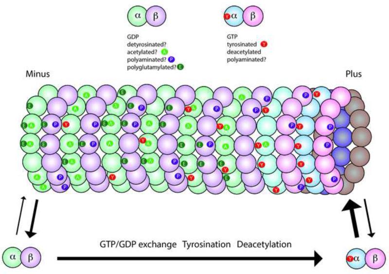
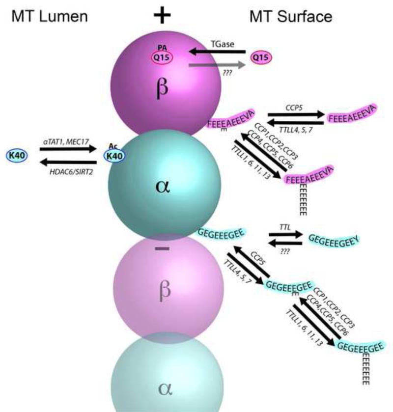
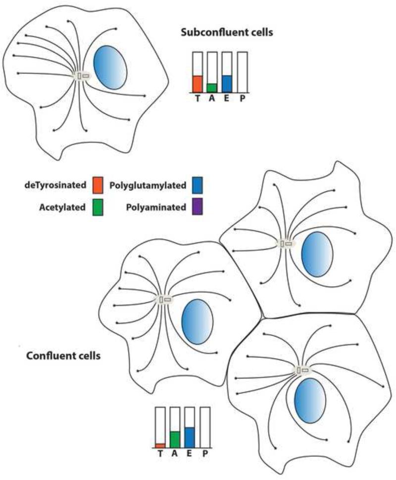
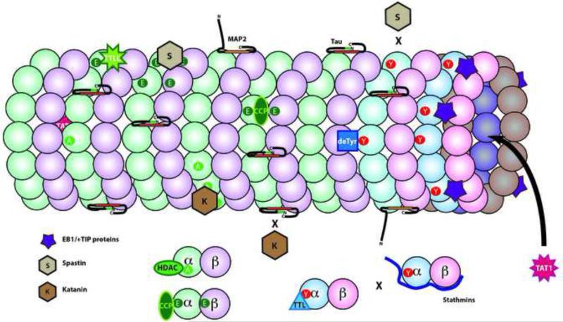
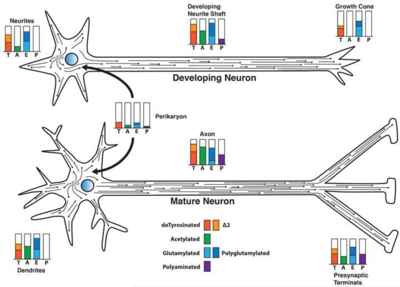
Tubulin and microtubules are subject to a remarkable number of post-translational modifications. Understanding the roles these modifications play in determining the functions and properties of microtubules has presented a major challenge that is only now being met. Many of these modifications are found concurrently, leading to considerable diversity in cellular microtubules, which varies with development, differentiation, cell compartment, and cell cycle. We now know that post-translational modifications of tubulin affect, not only the dynamics of the microtubules, but also their organization and interaction with other cellular components. Many early suggestions of how post-translational modifications affect microtubules have been replaced with new ideas and even new modifications as our understanding of cellular microtubule diversity comes into focus.

Keywords: acetylation; detyrosination; microtubule; polyamination; polyglutamylation; tubulin.


Figures

Figure 1
Figure 1. Microtubules and tubulins are subject to a variety of posttranslational modifications
This diagram illustrates the major modifications in axonal microtubules. Tubulin dimers are GTP-rich (blue/pink shading) in the soluble pool and on the plus end of the microtubule, but polymeric tubulin gradually hydrolyzes GTP and becomes GDP-rich (green/purple shading). Some modifications are associated specifically with tubulin polymerized in microtubules including acetylation (A), detyrosination, and polyglutamylation (E), while others may occur only on soluble tubulin like tyrosination (Y), or on either soluble or polymerized tubulin like polyamination (P). Some tubulins will have a single modification, while others have multiple modifications and some have no modification. However, the exact distribution along a microtubule and the fraction of the tubulin modified is highly variable, even within a single cell. A few rules have emerged such as tyrosinated tubulin is enriched at the plus ends of growing microtubules, while acetylation and detyrosination are more likely to be seen in the middle or minus end of the microtubule.
Figure 2
Figure 2. Pathways and sites for the major modifications of tubulin in a microtubule
Acetylation is primarily found on K40 of α-tubulins, which is exposed in the lumen of the microtubule. αTAT1 and Mec17 are the major enzymes responsible for acetylation (Ac), and HDAC6 and SIRT2 are the enzymes involved in deacetylation. The C-terminus of α-tubulin is a hot spot for modification and various combinations are illustrated. Tubulin Tyrosine Ligase (TTL) is the enzyme responsible for tyrosination of tubulin dimers, but the enzyme that catalyzes detyrosination remains to be identified. In contrast, multiple enzymes are capable of removing the penultimate glutamate residue to create Δ2-tubulin, including all six Cytoplasmic Carboxypeptidases (CCP1-6). These same enzymes can also trim the length of polyglutamylated chains, however, only one enzyme has been shown to remove the final iso-peptide linked glutamate (CCP5). Detyrosination is restricted to α-tubulins, but glutamylation and polyglutamylation can also occur on various glutamates in the C-terminal domain of β-tubulins. Multiple related enzymes have been identified that can add the iso-peptide link for initiating polyglutamylation (Tubulin Tyrosine Ligase Like family members (TTLL4,5,7)). Other members of the same family (TTLL1, 6, 11, 13) can add glycine to the same set of residues in the C-terminal domain of tubulins (not shown), but this reaction is seen only in ciliary microtubules. Curiously, the enzyme responsible for extension of polyglycine chains (TTLL10) is not functional in humans and only monoglycine modifications are seen. Finally, polyamination (PA) is mediated by transglutaminases, particularly TG2 in the nervous system. One putative modification site for polyamination identified by Mass Spectrometry is the conserved Q15 of β-tubulins. However, several other sites on both α- and β-tubulin may also be subject to polyamination. Enzyme(s) responsible for removing polyamines have not been confirmed and the degree to which polyamination is reversible in vivo is uncertain.
Figure 3
Figure 3. Posttranslational modifications of microtubules are dynamic and vary with cellular context
In some epithelial cells in culture, the degree of cell confluency and polarity of the cells affects the distribution and organization of modified microtubules. At early stages, microtubules are anchored to the microtubule organizing center at their negative ends. In subconfluent cells, microtubules are relatively rich in detyrosinated tubulin, but poor in acetylated tubulins. In confluent cells, the microtubule organization becomes more symmetrical and detryrosinated microtubules are reduced as acetylation of tubulin increases. Polyglutamylation levels appear to decrease slightly between these two states. Polyamination is generally undetectable in non-neuronal cells and may be specific to neurons.
Figure 4
Figure 4. Microtubule Associated Proteins and Posttranslational Modifications of Tubulins
One function of tubulin modifications is to affect the interaction of other proteins and enzymes. In complex with EB1, some +TIP binding proteins (purple 5 point star) bind selectively to tyrosinated tubulin. As a result, these +TIP proteins are enriched on the plus end of dynamic microtubules, which are rich in newly polymerized tyrosinated α-tubulin. Other proteins interact with particular tubulin isotypes like a subset of α-tubulins (deTyr for detyrosination) or to the C-termini of both α- and β-tubulins (TTLLs for polyglutamylation or polyglycylation). Tubulin acetylases (TAT1) are unique in binding to the interior of the microtubule lumen. Deacetylases (HDAC) or Tubulin Tyrosine Ligase (TTL) bind only to tubulin dimers. Stathmins bind to free tubulins as well, but partially overlap and compete with tubulin binding to TTL. The microtubule severing proteins, katanin (K) and spastin (S) are directed to particular microtubule domains by a combination of tubulin modifications and microtubule associated proteins. Katanin preferentially binds and severs acetylated domains of a microtubule, but katanin binding is inhibited by the presence of tau. In contrast, spastin preferentially binds and severs in polyglutamylated domains, but does not sever microtubules enriched in tyrosinated tubulin.
Figure 5
Figure 5. Posttranslational modifications of tubulin and microtubules vary in different regions of a neuron and change during neuronal differentiation
Microtubules in neuronal axons and dendrites are not anchored at the microtubule organizing center. The microtubules vary in length but have both plus and minus ends free. In the developing neuron (upper), when the axon is extending but the dendrites have not yet differentiated, the microtubules in all neurites are plus end out. Modification levels for detyrosination (T), Δ2 tubulin (Δ2), glutamylation (E), and polyamination (P) in the neuronal perikaryon are relatively low. Acetylation, glutamylation and detyrosination are elevated in the growing axon, but all three are reduced in the growth cone. Little or no acetylation is seen in the microtubules of the growth cone, consistent with the presence of highly dynamic microtubules. The minor neurites at this stage are lower in acetylation, but relatively rich in detyrosinated and glutamylated/polyglutamylated microtubules. Polyamination may be detectable in the growing axon at a low level. In a mature neuron (lower), as both dendrites and synaptic specializations form, tubulin modifications change both quantitatively and qualitatively. Dendritic microtubules exhibit mixed polarity as dendrites form, while axonal microtubules remain plus end out. Polyamination of axonal microtubules increases with maturation, but may be relatively low or absent from dendritic domains. Detyrosinated and Δ2 tubulin levels remain relatively high in differentiated dendrites and axons, while acetylation increases in both axon shafts and dendrites. Acetylation is also detectable in the distal axon and presynaptic regions, consistent with reduced numbers of highly dynamic microtubules in stable connections. During the differentiation and maturation of neurons, there are also changes in microtubule associated proteins in different neuronal subcellular domains. Adapted from Janke and Kneussel [5].

References

    1. Barra HS, et al. A soluble preparation from rat brain that incorporates into its own proteins ( 14 C)arginine by a ribonuclease-sensitive system and ( 14 C)tyrosine by a ribonuclease-insensitive system. J Neurochem. 1973;20:97–108. - PubMed
    1. Song Y, et al. Transglutaminase and Polyamination of Tubulin: Posttranslational Modification for Stabilizing Axonal Microtubules. Neuron. 2013;78:108–123. - PMC - PubMed
    1. Janke C, Bulinski JC. Post-translational regulation of the microtubule cytoskeleton: mechanisms and functions. Nat Rev Mol Cell Biol. 2011;12:773–786. - PubMed
    1. Wloga D, Gaertig J. Post-translational modifications of microtubules. J Cell Sci. 2010;123:3447–3455. - PMC - PubMed
    1. Janke C, Kneussel M. Tubulin post-translational modifications: encoding functions on the neuronal microtubule cytoskeleton. Trends Neurosci. 2010;33:362–372. - PubMed

Publication types