. 2015 Feb;359(2):521-536.
doi: 10.1007/s00441-014-2040-4. Epub 2014 Dec 3.

Expression of pluripotency factors in echinoderm regeneration

Affiliations

Expression of pluripotency factors in echinoderm regeneration

Vladimir S Mashanov et al. Cell Tissue Res. 2015 Feb.

Abstract

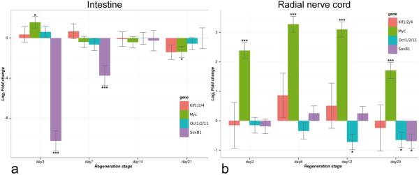
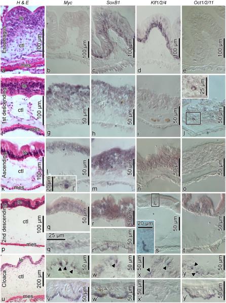
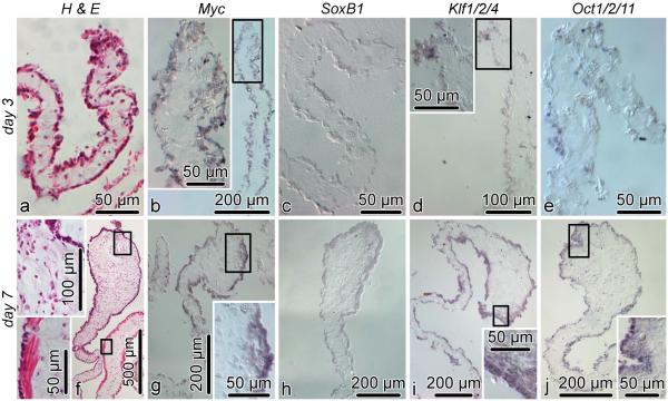
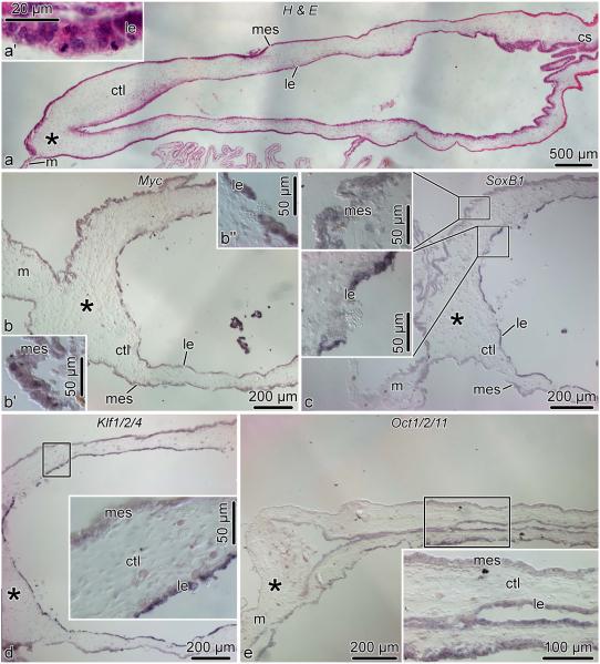
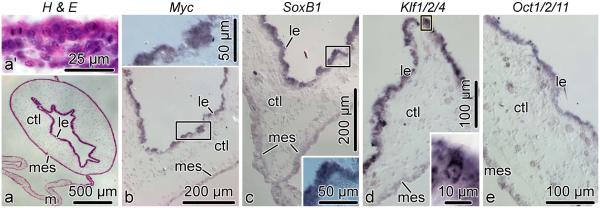
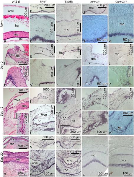
Cell dedifferentiation is an integral component of post-traumatic regeneration in echinoderms. As dedifferentiated cells become multipotent, we asked if this spontaneous broadening of developmental potential is associated with the action of the same pluripotency factors (known as Yamanaka factors) that were used to induce pluripotency in specialized mammalian cells. In this study, we investigate the expression of orthologs of the four Yamanaka factors in regeneration of two different organs, the radial nerve cord and the digestive tube, in the sea cucumber Holothuria glaberrima. All four pluripotency factors are expressed in uninjured animals, although their expression domains do not always overlap. In regeneration, the expression levels of the four genes were not regulated in a coordinated way, but instead showed different dynamics for individual genes and also were different between the radial nerve and the gut. SoxB1, the ortholog of the mammalian Sox2, was drastically downregulated in the regenerating intestine, suggesting that this factor is not required for dedifferentiation/regeneration in this organ. On the other hand, during the early post-injury stage, Myc, the sea cucumber ortholog of c-Myc, was significantly upregulated in both the intestine and the radial nerve cord and is therefore hypothesized to play a central role in dedifferentiation/regeneration of various tissue types.


Figures

Fig. 1
Fig. 1
Diagram showing domain organization for the predicted Oct1/2/11, Myc, Klf1/2/4, and SoxB1 proteins of the sea cucumber Holothuria glaberrima
Fig. 2
Fig. 2
Evolutionary relationships between Oct1/2/11 of H. glaberrima (arrowhead ) and homologous POU proteins in other animals. Green rectangle indicates class II of POU transcription factors. The numbers at branch nodes indicate the percentage of recovery of the branch in replicate trees in the bootstrap test. The tree is drawn to scale. The evolutionary distances were calculated using maximum composite likelihood method and are represented as number of base substitutions per site. The database accession numbers of the sequences used in the phylogenetic analysis are listed in Electronic Supplementary Material, Table S1
Fig. 3
Fig. 3
Evolutionary relationships between Myc of H. glaberrima (arrowhead ) and homologous proteins in other Deuterostomia
Fig. 4
Fig. 4
Evolutionary relationships between Klf1/2/4 of H. glaberrima (arrowhead ) and homologous proteins in other Deuterostomia. The tree was rooted using the Sp9 transcription factor of Danio rerio
Fig. 5
Fig. 5
Evolutionary relationships between SoxB1 of H. glaberrima (arrowhead) and Sox proteins of other animals. Green and red rectangles indicate the SoxB1 and SoxB2 subgroups of Sox transcription factors, respectively
Fig. 6
Fig. 6
Temporal expression pattern of Klf1/2/4, Myc, Oct1/2/11, and SoxB1 in the regenerating digestive tube (A) and radial nerve cord (B) as determined by quantitative real time RT-PCR. Three animals were used per time point. Expression values are plotted as fold change relative to the uninjured tissues in log2 scale. Error bars show standard deviation. P < 0.05,∗∗∗ P < 0.001. The full output of the MCMC.qpcr R package (Matz et al, 2013) containing relative expression values and corresponding p-values can be found in Electronic Supplementary Material, Text S3, and Text S4
Fig. 7
Fig. 7
Spatial expression of Myc, SoxB1, Klf1/2/4, and Oct1/2/11 in the non-eviscerated digestive tube. Each horizontal row of images corresponds to a different region of the digestive tube: a–e, esophagus; f–j, first descending intestine; k–o, ascending intestine; p–t, second descending intestine; u–y’, cloaca. The leftmost column of images (a, f, k, p, u) show reference paraffin sections (hematoxylin and eosin staining) for each of the regions of the digestive tube (a, esophagus; f, 1st descending intestine; k, ascending intesine; p, 2nd descending intestine; u, cloaca). The second thru fifth columns show in situ hybridization signal with riboprobes for each of the transcripts. All micrographs show transverse sections with the luminal epithelium to the top and the mesothelium to the bottom. In cases when two micrographs are shown for a particular gut region (l and l’; q and q’; r and r’, etc.), the upper micrograph (l, q, r, etc) shows the luminal epithelium and the bottom micrograph (l’, q’, r’, etc) shows the mesothelium. Insets in j and s show higher magnification views of the boxed areas. arrowheads indicate positively stained cells in the luminal epithelium of the cloaca. ctl, connective tissue layer; le, luminal epithelium; mes, mesothelium.
Fig. 8
Fig. 8
Spatial expression of Myc, SoxB1, Klf1/2/4, and Oct1/2/11 during early stages of visceral regeneration. The two horizontal rows of images (a thru e, f thru j) show cross sections of the regenerating intestine on day 3 and 7 post-evisceration, respectively. The first column (a and f ) shows hematoxylin and eosin stained reference sections. The second thru fifth columns (be, gj) show in situ hybridization labeling with respective riboprobes. Insets show high magnification view of the boxed areas in the respective main micrographs. ctl, connective tissue layer; mes, mesothelium.
Fig. 9
Fig. 9
Spatial expression of Myc, SoxB1, Klf1/2/4, and Oct1/2/11 in the regenerating intestine on day 14 post-evisceration. All micrographs show longitudinal sections of the regenerating intestine. The growing anterior tip of the gut rudiment (asterisk ) is on the left, whereas the cloacal stump (cs) is on the right. a, reference section stained with hematoxylin and eosin. a’, high-magnification view of the luminal epithelium of the growing intestinal rudiment showing abundant mitotic figures. b, Myc expression in the intestinal rudiment. b’ and b” show high-magnification views of the mesothelium and luminal epithelium, respectively. c, d, and e show expression of SoxB1, Klf1/2/4, and Oct1/2/11, respectively. Insets in c–e are high magnification views of the boxed areas in the respective main micrographs. cs, cloacal stump; ctl, connective tissue layer; le, luminal epithelium; m, mesentery; mes, mesothelium.
Fig. 10
Fig. 10
Expression of Myc, SoxB1, Klf1/2/4, and Oct1/2/11 in the tissues of the newly regenerated intestine on day 21 post-evisceration. All micrographs are cross sections. a, Low-magnification view of the newly regenerated intestine; reference paraffin section stained with hematoxylin and eosin. a’, high-magnification view of the luminal epithelium showing mitotic figures. b–e, In situ hybridization with riboprobes for Myc, SoxB1, Klf1/2/4, and Oct1/2/11, respectively. Insets show high magnification views of the boxed areas in the respective main micrographs. ctl, connective tissue layer; le, luminal epithelium; m, mesentery; mes, mesothelium
Fig. 11
Fig. 11
Expression of Myc, SoxB1, Klf1/2/4, and Oct1/2/11 in the uninjured and regenerating radial nerve cord. All sections are longitudinal. Horizontal rows of images correspond to the normal animals and three different stages of regeneration. The first vertical column shows reference paraffin sections stained with hematoxylin and eosin. The second thru fifth columns show in situ hybridization labeling with respective riboprobes. Insets show high magnification view of the boxed areas in the respective main micrographs. When two micrographs are shown for a particular condition (e.g., b and b’, the upper (e.g., b) and lower (e.g., b’) images show an overview and high-magnification view, respectively). ec, epineural canal; en, ectoneural neuroepithelium; hc, hyponeural canal; hn, hyponeural neuroepithelium; wvc, water-vacular canal; rnc, radial nerve cord.

References

    1. Ashton GH, Morton JP, Myant K, Phesse TJ, Ridgway RA, Marsh V, Wilkins JA, Athineos D, Muncan V, Kemp R, Neufeld K, Clevers H, Brunton V, Winton DJ, Wang X, Sears RC, Clarke AR, Frame MC, Sansom OJ. Focal adhesion kinase is required for intestinal regeneration and tumorigenesis downstream of Wnt/c-Myc signaling. Dev Cell. 2010;19(2):259–269. DOI 10.1016/j.devcel.2010.07.015, URL http://dx.doi.org/10.1016/j.devcel.2010.07.015. - DOI - PMC - PubMed
    1. Bhavsar RB, Tsonis PA. Exogenous Oct-4 inhibits lens transdifferentiation in the newt Notophthalmus viridescens. PLoS One. 2014;9(7):e102–510. DOI 0, URL http://dx.doi.org/0. - PMC - PubMed
    1. Brockes JP, Gates PB. Mechanisms underlying vertebrate limb regeneration: lessons from the salamander. Biochem Soc Trans. 2014;42(3):625–630. DOI 10.1042/BST20140002, URL http://dx.doi.org/10.1042/BST20140002. - DOI - PubMed
    1. Candelaria AG, Murray G, File SK, García-Arrarás JE. Contribution of mesenterial muscle dedifferentiation to intestine regeneration in the sea cucumber Holothuria glaberrima. Cell Tissue Res. 2006;325(1):55–65. DOI 10.1007/s00441-006-0170-z, URL http://dx.doi.org/10.1007/s00441-006-0170-z. - DOI - PubMed
    1. Christen B, Robles V, Raya M, Paramonov I, Izpisuá Belmonte JC. Regeneration and reprogramming compared. BMC Biol. 2010;8:5. DOI 10.1186/1741-7007-8-5, URL http://dx.doi.org/10.1186/1741-7007-8-5. - DOI - PMC - PubMed

Publication types

Substances

LinkOut - more resources