. 2014 Dec 2;3(6):e001274.
doi: 10.1161/JAHA.114.001274.

Loss of function in heparan sulfate elongation genes EXT1 and EXT 2 results in improved nitric oxide bioavailability and endothelial function

Affiliations

Loss of function in heparan sulfate elongation genes EXT1 and EXT 2 results in improved nitric oxide bioavailability and endothelial function

H L Mooij et al. J Am Heart Assoc. .

Abstract

Background: Heparanase is the major enzyme involved in degradation of endothelial heparan sulfates, which is associated with impaired endothelial nitric oxide synthesis. However, the effect of heparan sulfate chain length in relation to endothelial function and nitric oxide availability has never been investigated. We studied the effect of heterozygous mutations in heparan sulfate elongation genes EXT1 and EXT2 on endothelial function in vitro as well as in vivo.

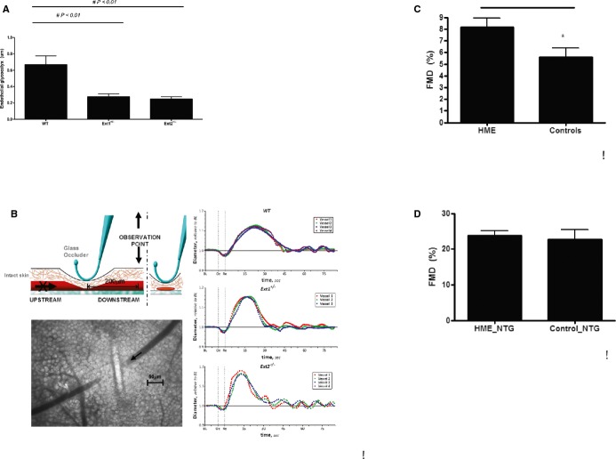
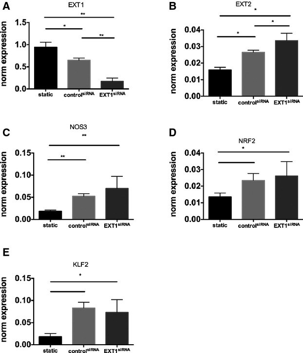
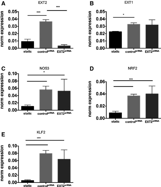
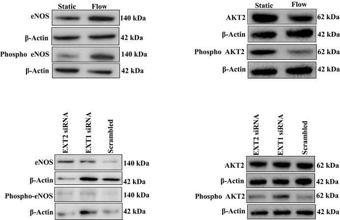
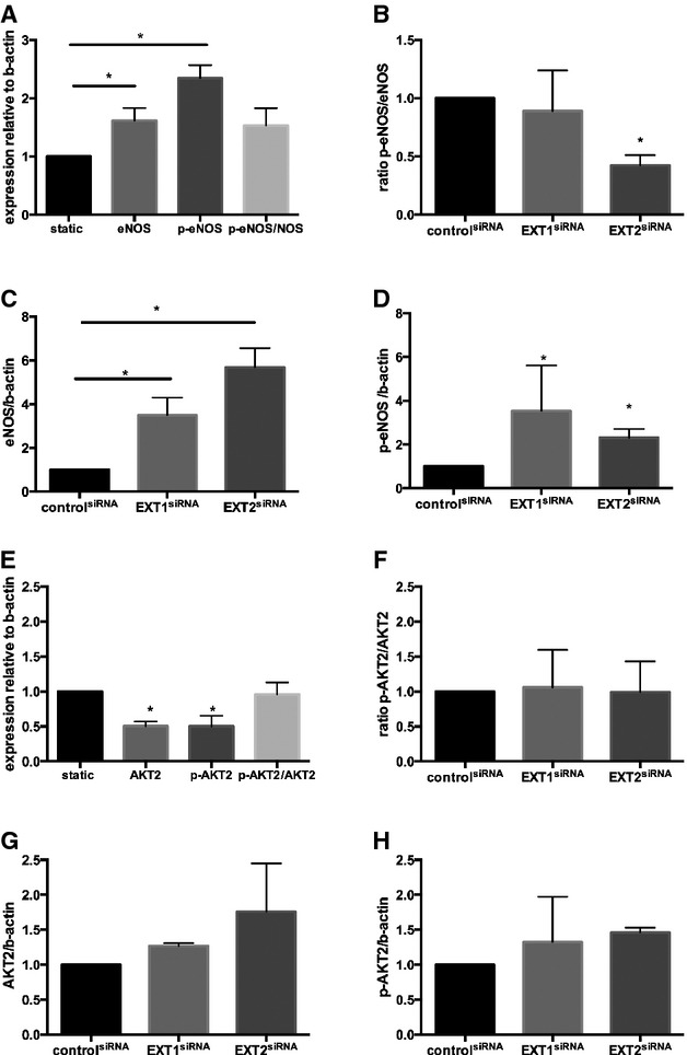
Methods and result: Flow-mediated dilation, a marker of nitric oxide bioavailability, was studied in Ext1(+/-) and Ext2(+/-) mice versus controls (n=7 per group), as well as in human subjects with heterozygous loss of function mutations in EXT1 and EXT2 (n=13 hereditary multiple exostoses and n=13 controls). Endothelial function was measured in microvascular endothelial cells under laminar flow with or without siRNA targeting EXT1 or EXT2. Endothelial glycocalyx and maximal arteriolar dilatation were significantly altered in Ext1(+/-) and Ext2(+/-) mice compared to wild-type littermates (glycocalyx: wild-type 0.67±0.1 μm, Ext1(+/-) 0.28±0.1 μm and Ext2(+/-) 0.25±0.1 μm, P<0.01, maximal arteriolar dilation during reperfusion: wild-type 11.3±1.0%), Ext1(+/-) 15.2±1.4% and Ext2(+/-) 13.8±1.6% P<0.05). In humans, brachial artery flow-mediated dilation was significantly increased in hereditary multiple exostoses patients (hereditary multiple exostoses 8.1±0.8% versus control 5.6±0.7%, P<0.05). In line, silencing of microvascular endothelial cell EXT1 and EXT2 under flow led to significant upregulation of endothelial nitric oxide synthesis and phospho-endothelial nitric oxide synthesis protein expression.

Conclusions: Our data implicate that heparan sulfate elongation genes EXT1 and EXT2 are involved in maintaining endothelial homeostasis, presumably via increased nitric oxide bioavailability.

Keywords: EXT; endothelial function; heparan sulfate; nitric oxide.


Figures

Figure 1.
Figure 1.
A, In vivo glycocalyx measurements in arterioles in WT mice, Ext1+/−, and Ext2+/− mice (n=7 mice per group). Values are presented as mean±SEM. Differences were analyzed using a nonparametric test (Mann–Whitney). *P<0.05. B, Graphic overview of single arteriolar vessel occlusion measurement in mice, and example of postdilation FMD curve in WT, Ext1+/−, and Ext2+/− mice as described in detail in the Methods section (n=4 to 6 mice per group analyzed). C, FMD of the brachial artery in controls (n=13) and in HME patients (n=13) with a heterozygous loss of function mutation in EXT1 or EXT2. D, Flow‐independent dilation of the brachial artery before and 4 minutes after administration on 0.4 mg nitroglycerine sublingually in controls (n=13) and in HME patients (n=13) with a heterozygous loss of function mutation in EXT1 or EXT2. Values are presented as mean±SEM. Differences were analyzed using a nonparametric test (Mann–Whitney). *P‐value<0.05. EXT indicates exostosin; FMD, flow‐mediated dilation; HME, hereditary multiple exostoses; NTG, nitroglycerine; WT, wild‐type.
Figure 2.
Figure 2.
EXT1 knockdown does not lead to further increase in NOS3 pathway. The effect of EXT1 knockdown on RNA expression of EXT1 (A), EXT2 (B), NOS3 (C), NRF2 (D), KLF2 (E) in HMVEC under laminar flow analyzed by RT‐PCR. EXT1 was silenced using siRNA against EXT1 as described. A scrambled siRNA was used as control. Flow induces a significant increase in NOS3, nuclear factor erythroid 2?related factor (NRF2) and kruppel like factor 2 (KLF2), which was not further increased by downregulation of EXT1. Data are from 4 independent experiments and presented as mean±SD. All silencing experiments were performed under laminar flow condition. Differences were analyzed using Student t test. *P<0.05; **P<0.01. EXT indicates exostosin; HMVEC, human microvascular endothelial cell; KLF2, kruppel like factor 2; NOS, nitric oxide synthesis; NRF‐2, nuclear factor erythroid 2?related factor; RT‐PCR, reverse‐transcription polymerase chain reaction.
Figure 3.
Figure 3.
EXT2 knockdown does not lead to further increase in NOS3 pathway. The effect of EXT2 knockdown on RNA expression of EXT2 (A), EXT2 (B), NOS3 (C), NRF2 (D), KLF2 (E) in HMVEC under laminar flow analyzed by RT‐PCR. EXT1 was silenced using siRNA against EXT2 as described. A scrambled siRNA was used as control. Flow induces a significant increase in NOS3, NRF2, and KLF2, which was not further increased by downregulation of EXT2. Data are from 4 independent experiments and presented as mean±SD. All silencing experiments were performed under laminar flow condition. Differences were analyzed using a nonparametric statistical test (Mann–Whitney). Differences were analyzed using Student t test. *P<0.05; ***P<0.005. EXT indicates exostosin; HMVEC, human microvascular endothelial cell; KLF2, kruppel like factor 2; NOS, nitric oxide synthesis; NRF‐2, nuclear factor erythroid 2?related factor; RT‐PCR, reverse‐transcription polymerase chain reaction.
Figure 4.
Figure 4.
Protein expression of eNOS, phospho‐eNOS, AKT and phospho‐AKT2 in HMVEC under static and laminar flow conditions. An example of eNOS, p‐eNOS, (left panel) AKT2 and p‐AKT2 protein expression (right panel) in HMVEC cell lysates under static and flow conditions with or without EXT1siRNA or EXT2siRNA. HMVECs were transfected with EXT1siRNA or EXT2siRNA, after which cells were studied under laminar flow. β‐Actin was used as loading control. eNOS indicates endothelial nitric oxide synthesis; EXT, exostosin; HMVEC, human microvascular endothelial cell.
Figure 5.
Figure 5.
Protein expression of eNOS, phospho‐eNOS, AKT, and phospho‐AKT2 in HMVEC under static and laminar flow conditions. A, The protein expression of eNOS, p‐eNOS, and the ratio of p‐eNOS/eNOS under flow as well as static condition. B, Effect of EXT1siRNA or EXT2siRNA on eNOS protein expression. C, Effect of EXT1siRNA or EXT2siRNA on phospho‐eNOS protein expression. D, Effect of EXT1siRNA or EXT2siRNA on the ratio of phospho‐eNOS/eNOS protein expression. All data are presented as mean±SEM (n=3 experiments). Differences between control and treatment groups were analyzed using nonparametric Kruskal–Wallis statistics. *P<0.05. E, Effect of flow on induction of AKT2 protein expression and phosphorylation status. F, Effect of EXT1siRNA or EXT2siRNA on AKT2 protein expression. G, Effect of EXT1siRNA or EXT2siRNA on phospho‐AKT2 protein expression. H, Effect of EXT1siRNA or EXT2siRNA on the ratio of phospho‐AKT2/AKT2 protein expression. Data are presented as mean±SEM for n=3 experiments. Differences between control and treatment groups were analyzed using nonparametric Kruskal–Wallis statistics. *P<0.05. eNOS indicates endothelial nitric oxide synthesis; EXT, exostosin; HMVEC, human microvascular endothelial cell.

References

    1. Murray CJ, Richards MA, Newton JN, Fenton KA, Anderson HR, Atkinson C, Bennett D, Bernabe E, Blencowe H, Bourne R, Braithwaite T, Brayne C, Bruce NG, Brugha TS, Burney P, Dherani M, Dolk H, Edmond K, Ezzati M, Flaxman AD, Fleming TD, Freedman G, Gunnell D, Hay RJ, Hutchings SJ, Ohno SL, Lozano R, Lyons RA, Marcenes W, Naghavi M, Newton CR, Pearce N, Pope D, Rushton L, Salomon JA, Shibuya K, Vos T, Wang H, Williams HC, Woolf AD, Lopez AD, Davis A. UK health performance: findings of the Global Burden of Disease Study 2010. Lancet. 2013; 381:997-1020. - PubMed
    1. Rudolph V, Freeman BA. Cardiovascular consequences when nitric oxide and lipid signaling converge. Circ Res. 2009; 105:511-522. - PMC - PubMed
    1. Flammer AJ, Anderson T, Celermajer DS, Creager MA, Deanfield J, Ganz P, Hamburg NM, Luscher TF, Shechter M, Taddei S, Vita JA, Lerman A. The assessment of endothelial function: from research into clinical practice. Circulation. 2012; 126:753-767. - PMC - PubMed
    1. Tarbell JM, Ebong EE. The endothelial glycocalyx: a mechano‐sensor and ‐transducer. Sci Signal. 2008; 1:pt8. - PubMed
    1. Mochizuki S, Vink H, Hiramatsu O, Kajita T, Shigeto F, Spaan JA, Kajiya F. Role of hyaluronic acid glycosaminoglycans in shear‐induced endothelium‐derived nitric oxide release. Am J Physiol Heart Circ Physiol. 2003; 285:H722-H726. - PubMed

Publication types

MeSH terms