. 2015 Jan 16;290(3):1348-63.
doi: 10.1074/jbc.M114.593830. Epub 2014 Dec 2.

Glycogen synthase kinase 3β orchestrates microtubule remodeling in compensatory glomerular adaptation to podocyte depletion

Affiliations

Glycogen synthase kinase 3β orchestrates microtubule remodeling in compensatory glomerular adaptation to podocyte depletion

Weiwei Xu et al. J Biol Chem. .

Abstract

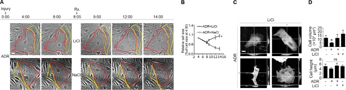
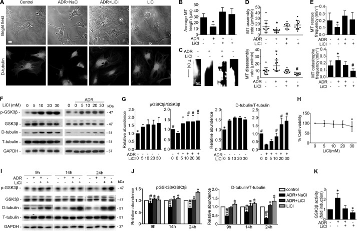
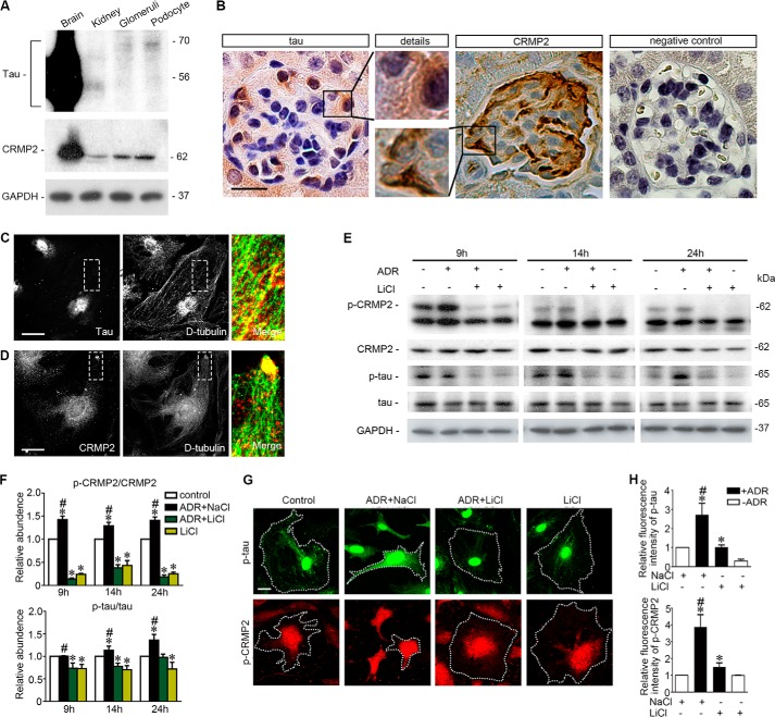
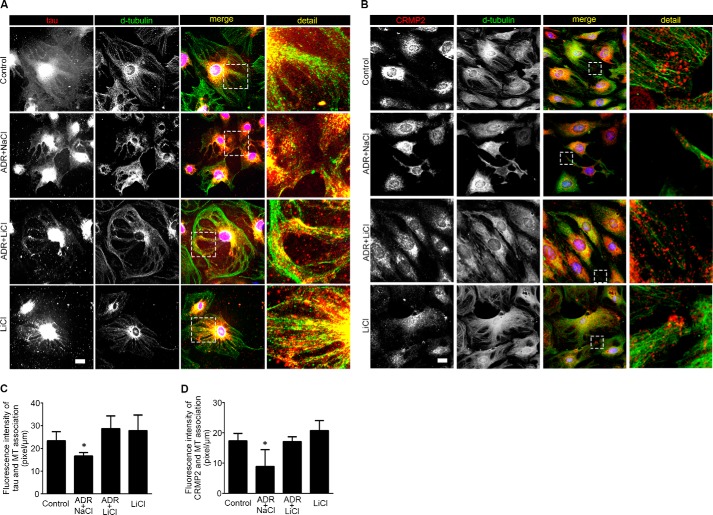
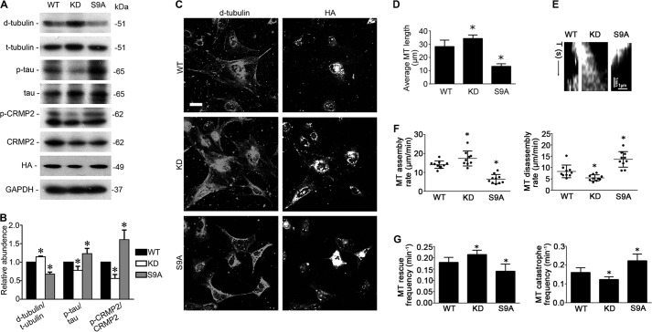
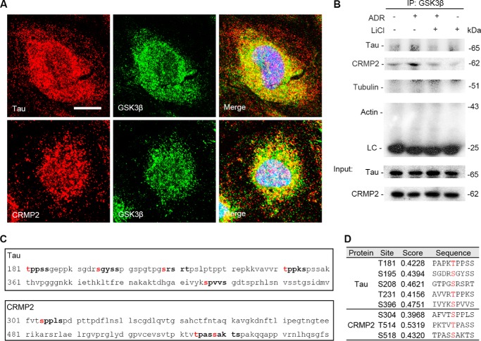
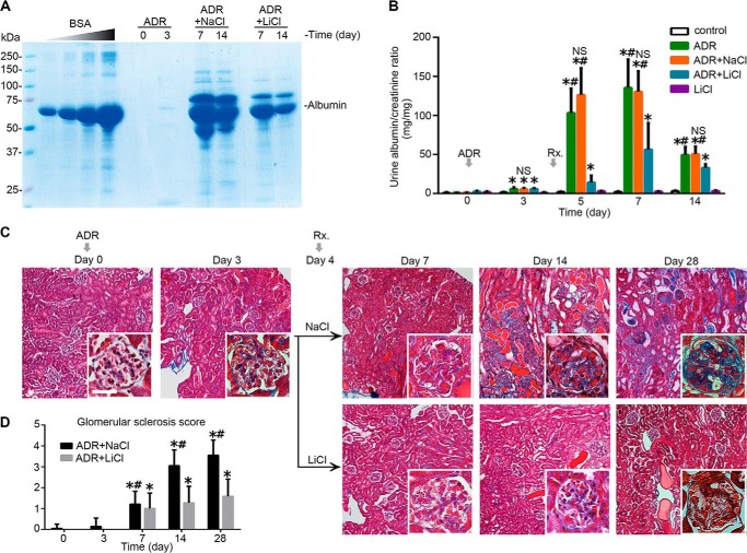
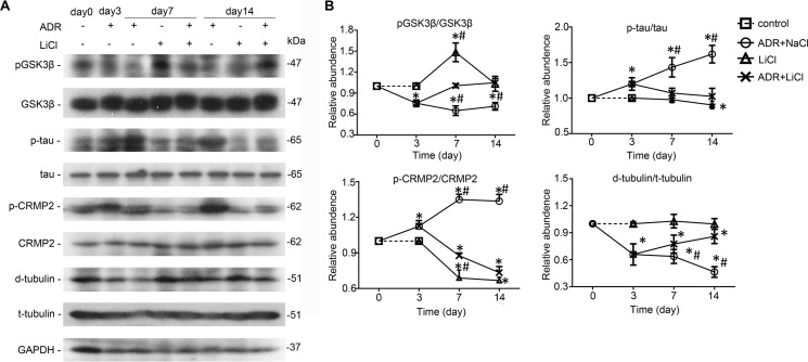
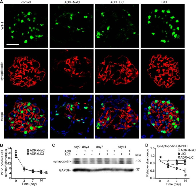
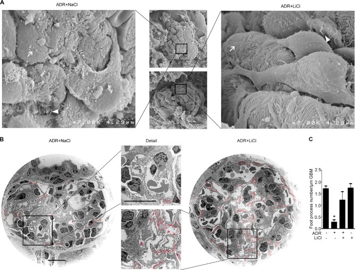
Reminiscent of neural repair, following podocyte depletion, remnant-surviving podocytes exhibit a considerable adaptive capacity to expand and cover the denuded renal glomerular basement membrane. Microtubules, one of the principal cytoskeletal components of podocyte major processes, play a crucial role in podocyte morphogenesis and podocyte process outgrowth, branching, and elongation. Here, we demonstrated that the microtubule-associated proteins Tau and collapsin response mediator protein (CRMP) 2, key regulators of microtubule dynamics, were abundantly expressed by glomerular podocytes in vivo and in vitro, interacted with glycogen synthase kinase (GSK)3β, and served as its putative substrates. GSK3β overactivity induced by adriamycin injury or by a constitutively active mutant of GSK3β augmented phosphorylation of Tau and CRMP2, concomitant with microtubule depolymerization, cell body shrinkage, and shortening of podocyte processes. Conversely, inhibition of GSK3β by a dominant negative mutant or by lithium, a Food and Drug Administration-approved neuroprotective mood stabilizer, diminished Tau and CRMP2 phosphorylation, resulting in microtubule polymerization, podocyte expansion, and lengthening of podocyte processes. In a mouse model of adriamycin-induced podocyte depletion and nephropathy, delayed administration of a single low dose of lithium attenuated proteinuria and ameliorated progressive glomerulosclerosis despite no correction of podocytopenia. Mechanistically, lithium therapy obliterated GSK3β overactivity, mitigated phosphorylation of Tau and CRMP2, and enhanced microtubule polymerization and stabilization in glomeruli in adriamycin-injured kidneys, associated with elongation of podocyte major processes. Collectively, our findings suggest that the GSK3β-dictated podocyte microtubule dynamics might serve as a novel therapeutic target to reinforce the compensatory glomerular adaptation to podocyte loss.

Keywords: Cytoskeleton; Glycogen Synthase Kinase 3β; Microtubule; Microtubule-associated Protein (MAP); Podocyte; Proteinuria; Tau Protein (Tau).


Figures

FIGURE 1.
FIGURE 1.
Rescue treatment with lithium chloride restores cellular shape in adriamycin-injured podocytes. A, time-lapse microscopy of differentiated and conditionally immortalized mouse podocytes following injury with adriamycin (ADR, 0.25 μg/ml) or vehicle. Lithium chloride (LiCl, 10 mm) or sodium chloride (NaCl, 10 mm) was added 8 h after ADR injury when cell shrinkage had occurred. Bar, 10 μm. B, morphometric quantification of cell sizes as relative to the original cell sizes at the 4th h (n = 30 cells per group). *, p < 0.05 versus sodium treatment at the same time point. C, podocytes were transfected to express GFP and then treated as stated in A. Representative micrographs of laser scanning confocal microscopy demonstrated the orthogonal views of podocytes at the 14th h following injury with adriamycin or vehicle. Bar, 10 μm. D, quantification of podocyte volume and height at the 14th h. *, p < 0.05 versus all other groups. ns, not significant.
FIGURE 2.
FIGURE 2.
Delayed treatment with lithium chloride improves microtubule dynamics and reinstates microtubule cytoskeletal integrity in adriamycin-injured podocytes. A, phase contrast fluorescent microscopy illustrated the distribution pattern of microtubule in podocytes treated with lithium chloride (LiCl, 10 mm) or sodium chloride (NaCl, 10 mm) 8 h after injury with vehicle or ADR (0.25 μg/ml). Micrographs were taken 14 h after ADR injury. Bar, 10 μm. B–E, podocytes were transfected to express GFP-tubulin and then treated as stated in A. Time-lapse fluorescence microscopy was performed 14 h following injury with adriamycin or vehicle to examine changes in the organization and dynamics of the GFP-labeled microtubules in live podocytes. B, quantification of microtubule (MT) length. *, p < 0.05 versus all other groups. C, kymographs of assembly and disassembly of single dynamic microtubules. D, quantification of microtubule assembly and disassembly rates. *, p < 0.05 versus all other groups; #, p < 0.05 versus control group. E, quantification of microtubule rescue and catastrophe frequency. *, p < 0.05 versus all other groups; #, p < 0.05 versus control group. F, differentiated podocytes were treated with different concentrations of LiCl 8 h after injury with ADR (0.25 μg/ml) or vehicle. Cells were collected 14 h after injury with ADR or vehicle, and cell lysates were prepared for immunoblot analysis for phosphorylated GSK3β (serine 9), total GSK3β, detyrosinated tubulin (D-tubulin), tyrosinated tubulin (T-tubulin), and GAPDH. G, arbitrary units of p-GSK3β/GSK3β ratios or detyrosinated tubulin/tyrosinated tubulin ratios expressed as immunoblot densitometric ratios as fold induction over the control group. *, p < 0.05 versus other groups; #, p < 0.05 versus ADR treatment only (n = 3 representative experiments). H, tetrazolium (MTT) assay of podocytes treated with different concentrations of LiCl. *, p < 0.05 versus the control group. I, after ADR (0.25 μg/ml) injury for 8 h, conditionally immortalized mouse podocytes were treated with LiCl (10 mm) or NaCl (10 mm) for 1, 6, and 16 h corresponding to 9, 14, and 24 h post-ADR injury. Cells were harvested at the indicated time, and cell lysates were subjected to immunoblot analysis for the indicated molecules. J, arbitrary units of p-GSK3β/GSK3β ratios or detyrosinated tubulin/tyrosinated tubulin ratios expressed as immunoblot densitometric ratios of the molecules as folds of the control group. *, p < 0.05 versus control group; #, p < 0.05 versus group ADR + LiCl (n = 3 representative experiments). K, GSK3β activity was assayed using the Tau (Ser(P)-396) phospho-ELISA kit, which measures the GSK3β-catalyzed phosphorylation of recombinant human Tau. The results were expressed as fold induction over the control group. *, p < 0.05 versus the control group (n = 4 representative experiments).
FIGURE 3.
FIGURE 3.
Microtubule-associated proteins Tau and CRMP2 are evidently expressed in podocytes in glomeruli, associated with microtubules, and regulated by adriamycin or lithium treatment. A, homogenates of mouse brain, kidney, isolated mouse glomeruli, and lysates of the differentiated mouse podocytes in culture were subjected to immunoblot analysis for Tau and CRMP2. Note that the long forms of Tau were probed as the major forms in isolated glomeruli and in cultured podocytes, whereas the short forms of Tau predominated in whole kidney homogenates. B, representative micrographs of peroxidase immunohistochemistry staining of mouse kidney for Tau and CRMP2 in glomeruli. Immunoperoxidase staining of Tau and CRMP2 was consistent with a pattern of podocyte-specific distribution. The primary antibody was replaced by nonimmune serum from the same species as a negative control, and no staining occurred. Bar, 20 μm. C, representative micrographs of dual color fluorescent immunocytochemistry staining of the differentiated mouse podocytes in culture indicated an association of Tau with stable microtubules as probed by detyrosinated tubulin (D-tubulin). Tau was closely associated with microtubule along the whole length. Bar, 10 μm. D, representative micrographs of dual color fluorescent immunocytochemistry staining of the differentiated mouse podocytes in culture indicated an association of CRMP2 with stable microtubules as probed by detyrosinated tubulin (D-tubulin). CRMP2 was associated with microtubules and clustered at the leading edge of podocyte extensions. Bar, 10 μm. E, after ADR (0.25 μg/ml) or vehicle treatment for 8 h, conditionally immortalized mouse podocytes were treated with or without lithium chloride (10 mm) or sodium chloride (10 mm) for 1, 6, and 16 h corresponding to 9, 14, and 24 h post-ADR injury. Cells were harvested at the indicated time points, and cell lysates were subjected to immunoblot analysis for the indicated proteins. F, arbitrary units of p-CRMP2/CRMP2 ratios or p-Tau/Tau ratios expressed as immunoblot densitometric ratios of the molecules as folds of the control group. *, p < 0.05 versus control group; #, p < 0.05 versus ADR + LiCl group, (n = 3 representative experiments). G, representative micrographs of fluorescent immunocytochemistry staining of phosphorylated Tau and phosphorylated CRMP2 in podocytes 14 h following different treatments as stated in Fig. 2A. Bar, 5 μm. H, morphometric analysis of relative fluorescence intensity of p-Tau and p-CRMP2 in podocytes. *, p < 0.05 versus control group; #, p < 0.05 versus ADR + LiCl group; (n = 6).
FIGURE 4.
FIGURE 4.
Lithium treatment reinstates the association of Tau and CRMP2 with microtubules and improves microtubule integrity in adriamycin-injured podocytes. A, differentiated podocytes were treated as stated in Fig. 2A and prepared at 14 h for immunocytochemistry staining. Representative micrographs of dual color fluorescent immunocytochemistry staining of detyrosinated tubulin and Tau. Tau was associated with microtubules in control and lithium (LiCl)-treated podocytes. ADR injury induced podocyte shrinkage, disrupted microtubule integrity, and lessened the association between Tau and microtubule. The effect of ADR was abrogated by rescue treatment with LiCl. Sodium treatment (NaCl) served as a control for LiCl treatment. Bar, 10 μm. B, representative micrographs of dual color fluorescent immunocytochemistry staining of detyrosinated tubulin (D-tubulin) and CRMP2 in podocytes. There were some clusters of CRMP2 at the leading edges in control and LiCl-treated cells. Adriamycin injury reduced podocyte sizes, disrupted microtubule networks, and diminished CRMP2 clusters at the leading edges of cell projections. Rescue treatment with lithium chloride restored cell shape and reinstated CRMP2 clustering at the leading edges of podocytes. Bar, 10 μm. C, quantitative morphometric analysis of the fluorescence intensity of microtubule-associated Tau per unit length of microtubules. *, p < 0.05 versus all other groups (n = 3 representative experiments). D, quantitative morphometric analysis of the fluorescence intensity of microtubule-associated CRMP2 per unit length of microtubule. *, p < 0.05 versus all other groups (n = 3 representative experiments).
FIGURE 5.
FIGURE 5.
GSK3β controls microtubule dynamics by regulating Tau and CRMP2. A, differentiated podocytes were transfected with vectors encoding the hemagglutinin (HA)-conjugated wild type GSK3β (WT), kinase-dead mutant of GSK3β (KD), or constitutively active mutant of GSK3 β (S9A). Cells were collected 48 h after transfection and prepared for immunoblot analysis or fluorescent immunocytochemistry staining. B, densitometric analysis of Western immunoblot in A. *, p < 0.05 versus all other groups (n = 3). C, representative micrographs of dual color fluorescent immunocytochemistry staining of detyrosinated tubulin (D-tubulin) and HA. Bar, 10 μm. D–G, differentiated podocytes were transfected to express both GFP-tubulin and WT, KD, or S9A mutant of GSK3β. Time-lapse fluorescence microscopy of live podocytes was performed 48 h after transfection, and micrographs were recorded and analyzed. D, quantification of microtubule (MT) length. *, p < 0.05 versus all other groups (n = 3). E, kymographs of assembly and disassembly of single dynamic microtubules. F, quantification of microtubule assembly and disassembly rate. *, p < 0.05 versus all other groups (n = 3). G, quantification of microtubule rescue and catastrophe frequency. *, p < 0.05 versus all other groups; (n = 3).
FIGURE 6.
FIGURE 6.
Tau and CRMP2 colocalize and physically interact with GSK3β as its putative substrates in podocytes. A, representative micrographs of laser scanning confocal microscopy of dual color fluorescent immunocytochemistry staining of GSK3β and Tau or CRMP2 in differentiated conditionally immortalized mouse podocytes. Evident colocalization of Tau or CRMP2 with GSK3β was noted in the cytoplasm of differentiated podocytes. Bar, 5 μm. B, podocytes were treated as described in Fig. 2A, and cell lysates were prepared at 14 h for immunoprecipitation followed by immunoblot analysis for indicated molecules. LC, IgG light chain. An aliquot of cell lysates from each group served as input control. C, in silico analysis indicated that amino acid residues Thr-181, Ser-195, Ser-208, Thr-231, and Ser-396 of the longest isoform of small Tau and amino acid residues Ser-304, Thr-514, and Ser-518 of CRMP2 reside in the consensus motifs for phosphorylation by GSK3β. D, characteristics of consensus GSK3β phosphorylation motifs in Tau and CRMP2, including the predicted phosphorylation sites, prediction confidence scores, and sequences, as estimated by in silico analysis.
FIGURE 7.
FIGURE 7.
Delayed administration of a single low dose of lithium induces proteinuria remission and ameliorates glomerulosclerosis in experimental adriamycin nephropathy. Mice were subjected to ADR injury (10 mg/kg, tail vein injection) on day 0 and received a single intraperitoneal injection of lithium chloride (40 mg/kg) or an equal molar amount (1 meq/kg) of sodium chloride (NaCl) as saline on day 4. A, urine was collected at the indicated time points and subjected to SDS-PAGE followed by Coomassie Brilliant Blue staining. Bovine serum albumin (BSA) of 5, 10, 20, and 40 μg served as standard controls. Urine (1.5 μl) from each group was loaded. B, quantification of urine albumin levels adjusted for urine creatinine concentrations. *, p < 0.05 versus control group; #, p < 0.05 versus group ADR + LiCl. (n = 6). C, representative micrographs demonstrating Masson's trichrome staining of mouse kidneys. Bars, 20 μm. D, morphometric analysis of glomerulosclerosis score. *, p < 0.05 versus day 0; #, p < 0.05 versus group ADR + LiCl (n = 6).
FIGURE 8.
FIGURE 8.
Lithium counteracts GSK3β overactivity, diminishes Tau and CRMP2 phosphorylation, and reinstates microtubule integrity in adriamycin-injured glomeruli. A, immunoblot analysis of homogenates of isolated glomeruli for indicated molecules. B, arbitrary units of p-GSK3β/GSK3β ratios, p-Tau/Tau ratios, p-CRMP2/CRMP2 ratios, and detyrosinated tubulin (d-tubulin)/tyrosinated tubulin (t-tubulin) ratios expressed as immunoblot densitometric ratios of the molecules as folds of the control groups. *, p < 0.05 versus day 0; #, p < 0.05 versus group ADR + LiCl (n = 6).
FIGURE 9.
FIGURE 9.
Delayed lithium therapy potentiates glomerular adaptation to podocyte depletion in adriamycin nephropathy. A, fluorescence immunohistochemistry staining of kidney specimens procured on day 14 for synaptopodin and WT-1. Lithium chloride therapy abrogated the ADR-diminished synaptopodin expression in the glomerulus but barely improved the adriamycin-mitigated WT-1 expression. Bar, 30 μm. B, absolute count of WT-1-positive podocytes per glomerulus on kidney specimens procured from mice on days 0, 3, 7, and 14. *, p < 0.05 versus other time points, NS, not significant versus group ADR + LiCl (n = 6). C, Western immunoblot analysis of homogenates of isolated glomeruli for synaptopodin. D, densitometric analysis of the immunoblots of synaptopodin. *, p < 0.05 versus ADR-treated groups; #, p < 0.05 versus group ADR + LiCl (n = 6).
FIGURE 10.
FIGURE 10.
Rescue treatment with a single low dose of lithium, an inhibitor of GSK3β and Food and Drug Administration-approved mood stabilizer, in experimental adriamycin nephropathy promotes major process elongation in remnant surviving podocytes to compensate for the loss of podocytes and to cover the denuded glomerular basement membrane. Kidney cortical tissues were procured on day 7 from ADR-injured mice receiving rescue treatment of lithium chloride (40 mg/kg) or an equal molar amount of sodium chloride as saline. Kidney specimens were processed from scanning or transmission electron microscopy. A, scanning electron microscopy of glomeruli. White arrows indicate podocyte foot process effacement; white arrowheads indicate podocyte microvillous transformation. B, transmission electron microscopy of glomeruli. Major processes were outlined by red lines for the purpose of stereological analysis to estimate the lengths of podocyte major processes. Bar, 10 μm. C, absolute count of the number of foot processes per unit length of glomerular basement membrane as evaluated by transmission electron microscopy. *, p < 0.05 versus all other groups.

Similar articles

Cited by

References

    1. Fukuda A., Chowdhury M. A., Venkatareddy M. P., Wang S. Q., Nishizono R., Suzuki T., Wickman L. T., Wiggins J. E., Muchayi T., Fingar D., Shedden K. A., Inoki K., Wiggins R. C. (2012) Growth-dependent podocyte failure causes glomerulosclerosis. J. Am. Soc. Nephrol. 23, 1351–1363 - PMC - PubMed
    1. Kriz W. (2012) Glomerular diseases: podocyte hypertrophy mismatch and glomerular disease. Nat. Rev. Nephrol. 8, 618–619 - PubMed
    1. D'Agati V. D., Kaskel F. J., Falk R. J. (2011) Focal segmental glomerulosclerosis. N. Engl. J. Med. 365, 2398–2411 - PubMed
    1. D'Agati V. D. (2012) Pathobiology of focal segmental glomerulosclerosis: new developments. Curr. Opin. Nephrol. Hypertens. 21, 243–250 - PubMed
    1. Wharram B. L., Goyal M., Wiggins J. E., Sanden S. K., Hussain S., Filipiak W. E., Saunders T. L., Dysko R. C., Kohno K., Holzman L. B., Wiggins R. C. (2005) Podocyte depletion causes glomerulosclerosis: diphtheria toxin-induced podocyte depletion in rats expressing human diphtheria toxin receptor transgene. J. Am. Soc. Nephrol. 16, 2941–2952 - PubMed

Publication types

MeSH terms

LinkOut - more resources