. 2014 Dec 16;111(50):17715-22.
doi: 10.1073/pnas.1418796111. Epub 2014 Dec 2.

Microtubule nucleation remote from centrosomes may explain how asters span large cells

Affiliations

Microtubule nucleation remote from centrosomes may explain how asters span large cells

Keisuke Ishihara et al. Proc Natl Acad Sci U S A. .

Abstract

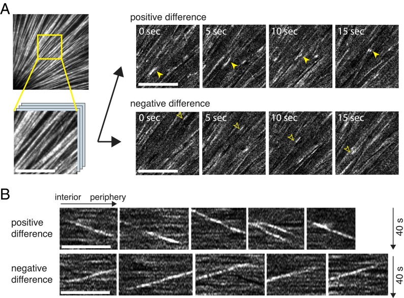
A major challenge in cell biology is to understand how nanometer-sized molecules can organize micrometer-sized cells in space and time. One solution in many animal cells is a radial array of microtubules called an aster, which is nucleated by a central organizing center and spans the entire cytoplasm. Frog (here Xenopus laevis) embryos are more than 1 mm in diameter and divide with a defined geometry every 30 min. Like smaller cells, they are organized by asters, which grow, interact, and move to precisely position the cleavage planes. It has been unclear whether asters grow to fill the enormous egg by the same mechanism used in smaller somatic cells, or whether special mechanisms are required. We addressed this question by imaging growing asters in a cell-free system derived from eggs, where asters grew to hundreds of microns in diameter. By tracking marks on the lattice, we found that microtubules could slide outward, but this was not essential for rapid aster growth. Polymer treadmilling did not occur. By measuring the number and positions of microtubule ends over time, we found that most microtubules were nucleated away from the centrosome and that interphase egg cytoplasm supported spontaneous nucleation after a time lag. We propose that aster growth is initiated by centrosomes but that asters grow by propagating a wave of microtubule nucleation stimulated by the presence of preexisting microtubules.

Keywords: Xenopus; aster; centrosome; embryo; microtubule nucleation.


Conflict of interest statement

The authors declare no conflict of interest.

Figures

Fig. 1.
Fig. 1.
Reconstitution of large microtubule asters in a cell-free system. (A) High density of beads coated with AurkA antibody in interphase Xenopus egg extract nucleate many asters that mutually inhibit their growth upon contact, leading to limited radii; 34 min after calcium addition. (B) At low bead densities, asters are spatially separated and grow in an uninterrupted manner. Large asters spanning ∼500 µm in radius are observed; 45 min after calcium addition. Wide-field images of fluorescently labeled tubulin. (C) Asters grow and eventually span the entire cytoplasm of Xenopus laevis zygote. Laser scanning confocal images of eggs fixed at 45 min after fertilization and stained for tubulin by immunofluorescence.
Fig. 2.
Fig. 2.
Large asters assemble in the absence of dynein-mediated microtubule outward sliding. (A) Asters assembled between uncoated coverslips show curved and disorganized microtubules. Image acquired on a wide-field microscope; 15 min after calcium addition. (Scale bar, 50 µm.) (B) First frame of a fluorescence speckle microscopy movie (Movie S1) showing labeled tubulin in an aster near its center. Image acquired on a spinning disk microscope. Movies were subjected to particle image velocimetry to determine the outward sliding rate as 6.1 ± 2.8 µm/min (SD, n = 12 asters). (Scale bar, 10 µm.) (B’) Kymographs generated from yellow line in B, where left to right corresponds to interior to periphery of the aster. (Scale bar, 10 µm.) (C–D’) Same as A–B’, in the presence of p150-CC1 protein to inhibit dynein motor activity. Note the decreased curving of aster microtubules and the complete suppression of microtubule outward sliding, −0.16 ± 0.25 µm/min (n = 12 asters) (Movie S2). (E–F’) Same as A–B’, with the coverslip surface passivated by PLL-PEG. This also resulted in the decrease of microtubule curving and the reduction of microtubule outward sliding to 2.7 ± 1.2 µm/min (n = 6 asters) (Movie S3).
Fig. 3.
Fig. 3.
Tubulin intensity difference imaging reveals dynamic microtubule plus-ends in a growing aster. (A) Microtubules were imaged with labeled tubulin by TIRF microscopy (Movie S4). To facilitate analysis of polymer dynamics, intensity differences of subsequent images were calculated to yield maps of polymerization (positive difference; Movie S5) and depolymerization (negative difference; Movie S6) (Materials and Methods) (Scale bars, 10 µm.) (B) Kymographs were constructed to measure the rates of polymerization and depolymerization observed above. Polymerization proceeded outward at 30.0 ± 4.3 µm/min (SD, n = 56 ends from four asters), whereas depolymerization traveled inward at 41.8 ± 5.2 µm/min (SD, n = 50 ends from four asters), representing plus-end dynamics. (Scale bars, 10 µm.)
Fig. 4.
Fig. 4.
Quantification of microtubule plus-ends during aster growth. (A) Live imaging of labeled tubulin and the plus-end tracking protein EB1-GFP using wide-field microscopy. Images show dynein-inhibited asters assembled under PLL-PEG passivated coverslips. (Insets) Magnification of region in yellow box. Time indicates time after calcium addition. (Scale bars, 100 µm.) (B) Time evolution of the number of EB1 comets detected at a given distance. Comets were detected and tracked using the PlusTipTracker software (19) (Materials and Methods). (Inset) Half-maximum positions of the EB1 comet number at different time points. (C) Time evolution of the EB1 comet density at a given distance. (D) Gallery showing the EB1 density of multiple examples of aster growth. Curves in each plot are 2 min apart.
Fig. 5.
Fig. 5.
Microtubules in the aster appear at a distance from the organizing center. (A) Maximum intensity projection of four consecutive EB1-GFP images taken over 9.6 s. Same aster as in Fig. 4A at 28 min after calcium addition. (A’) Particle tracking was applied to the same four images (shown in A) to measure microtubule nucleation from AurkA bead (red asterisk). All EB1 comets detected are represented as dots varying from green to blue (frame 1–4). Blue lines represent all detected tracks. Red circles indicate tracks that were detected as crossing the 40-µm perimeter (line). Details in Materials and Methods. (B) AurkA bead nucleation rate measure as in A’. For each time point, number of comets crossing over radii of 38, 39, 40, 41, and 42 µm are shown in the plot. (C) Total number of EB1 comets increase dramatically as the aster grows (blue dots). The predicted number of EB1 comets based on the nucleation of AurkA bead alone (red plus signs) is significantly lower. (Inset) Ratio of bead (red) to total (blue) at each time point.
Fig. 6.
Fig. 6.
Microtubules assemble in the interphase cytoplasm in the absence of centrosomes. (A) Time lapse image of spontaneously nucleated microtubules in the interphase extract with EB1-GFP. Time denotes time after calcium addition. (B) Quantification of EB1 comet density over time for the same reaction as in A. Different colors represent different positions of the same coverslip. (C) Tetrahymena pellicles in interphase extract nucleate microtubules and form large radial arrays of microtubules resembling asters. (Scale bar, 100 µm.) (D) Immunofluorescence image showing the pattern of tubulin in eggs fixed at 90 min after electroactivation. (Scale bar, 200 µm.)
Fig. 7.
Fig. 7.
A two-step model for large aster growth. At early time points, aster growth is initiated by AurkA-dependent microtubule nucleation at the centrosome. Later in interphase, aster growth is sustained by microtubule nucleation away from the centrosome, presumably stimulated by preexisting microtubules.

References

    1. Wilson EB. The Cell in Development and Inheritance. Macmillan; New York: 1925.
    1. Wühr M, et al. Evidence for an upper limit to mitotic spindle length. Curr Biol. 2008;18(16):1256–1261. - PMC - PubMed
    1. Wühr M, Tan ES, Parker SK, Detrich HW, 3rd, Mitchison TJ. A model for cleavage plane determination in early amphibian and fish embryos. Curr Biol. 2010;20(22):2040–2045. - PMC - PubMed
    1. Wühr M, Dumont S, Groen AC, Needleman DJ, Mitchison TJ. How does a millimeter-sized cell find its center? Cell Cycle. 2009;8(8):1115–1121. - PMC - PubMed
    1. Nguyen PA, Groen AC, et al. Spatial organization of cytokinesis signaling reconstituted in a cell-free system. Science. 2014;346(6206):244–247. - PMC - PubMed

Publication types

LinkOut - more resources