

Contrast-enhanced spectral mammography: comparison with conventional mammography and histopathology in 152 women
- PMID: 25469079
- PMCID: PMC4248623
- DOI: 10.3348/kjr.2014.15.6.689
Contrast-enhanced spectral mammography: comparison with conventional mammography and histopathology in 152 women
Abstract
Objective: The goal of the study was to compare conventional mammography (MG) and contrast-enhanced spectral mammography (CESM) in preoperative women.
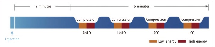
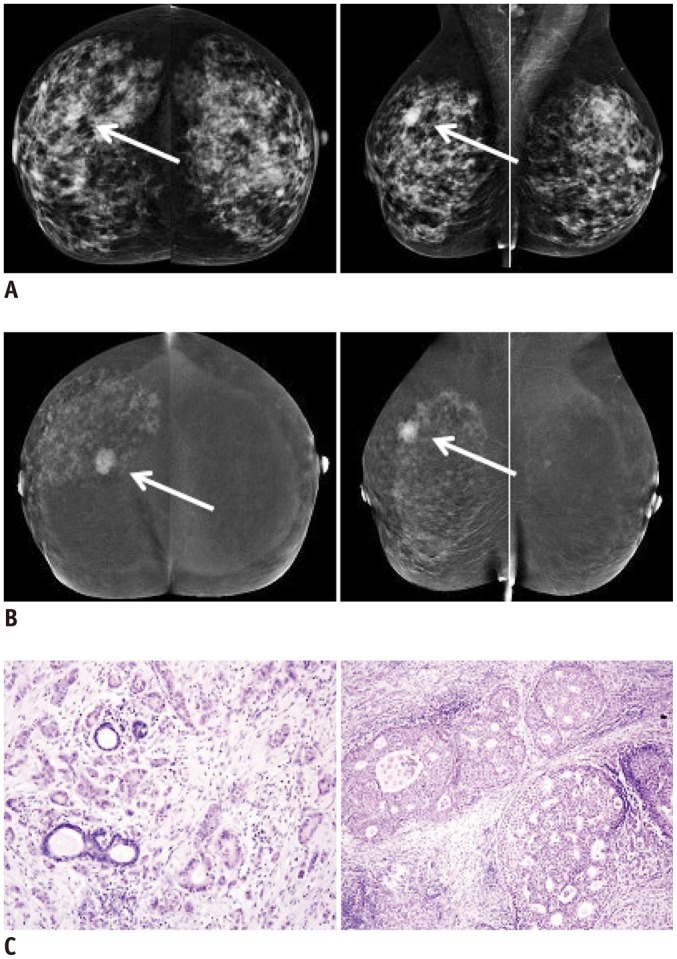
Materials and methods: The study was approved by the local Ethics Committee and all participants provided informed consent. The study included 152 consecutive patients with 173 breast lesions diagnosed on MG or CESM. All MG examinations and consults were conducted in one oncology centre. Non-ionic contrast agent, at a total dose of 1.5 mL/kg body weight, was injected intravenous. Subsequently, CESM exams were performed with a mammography device, allowing dual-energy acquisitions. The entire procedure was done within the oncology centre. Images from low and high energy exposures were processed together and the combination provided an "iodine" image which outlined contrast up-take in the breast.
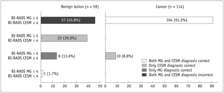
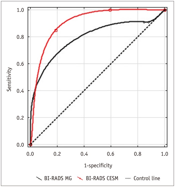
Results: MG detected 157 lesions in 150 patients, including 92 infiltrating cancers, 12 non-infiltrating cancers, and 53 benign lesions. CESM detected 149 lesions in 128 patients, including 101 infiltrating cancers, 13 non-infiltrating cancers, and 35 benign lesions. CESM sensitivity was 100% (vs. 91% for MG), specificity was 41% (vs. 15% for MG), area under the receiver operating characteristic curve was 0.86 (vs. 0.67 for MG), and accuracy was 80% (vs. 65% for MG) for the diagnosis of breast cancer. Both MG and CESM overestimated lesion sizes compared to histopathology (p < 0.001).
Conclusion: CESM may provide higher sensitivity for breast cancer detection and greater diagnostic accuracy than conventional mammography.
Keywords: Breast cancer; Contrast enhanced spectral mammography; Mammography.
Figures
References
-
- Jochelson M. Advanced imaging techniques for the detection of breast cancer. Am Soc Clin Oncol Educ Book. 2012:65–69. - PubMed
-
- Burhenne HJ, Burhenne LW, Goldberg F, Hislop TG, Worth AJ, Rebbeck PM, et al. Interval breast cancers in the Screening Mammography Program of British Columbia: analysis and classification. AJR Am J Roentgenol. 1994;162:1067–1071. discussion 1072-1075. - PubMed
-
- Robertson CL. A private breast imaging practice: medical audit of 25,788 screening and 1,077 diagnostic examinations. Radiology. 1993;187:75–79. - PubMed
-
- Mandelson MT, Oestreicher N, Porter PL, White D, Finder CA, Taplin SH, et al. Breast density as a predictor of mammographic detection: comparison of interval- and screen-detected cancers. J Natl Cancer Inst. 2000;92:1081–1087. - PubMed
-
- Kolb TM, Lichy J, Newhouse JH. Comparison of the performance of screening mammography, physical examination, and breast US and evaluation of factors that influence them: an analysis of 27,825 patient evaluations. Radiology. 2002;225:165–175. - PubMed
Publication types
MeSH terms
Substances
LinkOut - more resources
Full Text Sources
Other Literature Sources
Medical
