

Review
doi: 10.3348/kjr.2014.15.6.724.
Epub 2014 Nov 7.
Lymphangiography to treat postoperative lymphatic leakage: a technical review
Affiliations
- PMID: 25469083
- PMCID: PMC4248627
- DOI: 10.3348/kjr.2014.15.6.724
Item in Clipboard
Review
Lymphangiography to treat postoperative lymphatic leakage: a technical review
Korean J Radiol.
2014 Nov-Dec.
Abstract
In addition to imaging the lymphatics and detecting various types of lymphatic leakage, lymphangiography is a therapeutic option for patients with chylothorax, chylous ascites, and lymphatic fistula. Percutaneous thoracic duct embolization, transabdominal catheterization of the cisterna chyli or thoracic duct, and subsequent embolization of the thoracic duct is an alternative to surgical ligation of the thoracic duct. In this pictorial review, we present the detailed technique, clinical applications, and complications of lymphangiography and thoracic duct embolization.
Keywords: Chylothorax; Chylous ascites; Lymphangiography; Lymphatic fistula.
Figures
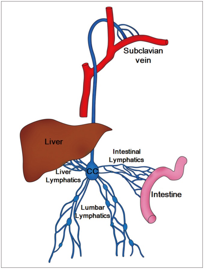
Schematic drawing of chyle pathway. Lumbar lymphatics drain not only lumbar region but also lower extremities. Lumbar, intestinal, and liver lymphatics join and form cisterna chyli (CC) which finally empties into left subclavian vein.
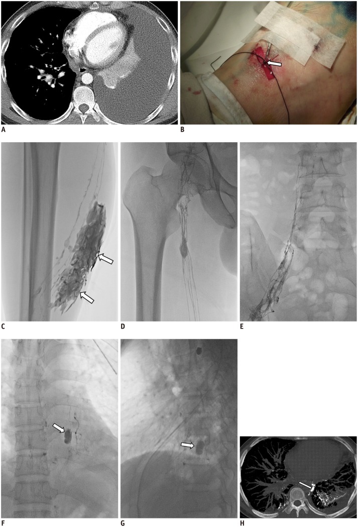
35-year-old man with left chylothorax after lung biopsy using video-assisted thoracoscopy. A. Axial CT scan obtained one week prior to LG shows large amount of left pleural effusion (chylothorax). B. Isolated lymphatic of dorsum of right foot was cannulated using 30-G LG needle and both needle and lymphatic were firmly tied (arrow). C. Radiographic image obtained 5 minutes following Lipiodol injection shows Lipiodol extravasation (arrows) at calf level. D, E. Radiographic images obtained 15 minutes following Lipiodol injection show good opacification of inguinal and pelvic lymph nodes as well as ascending lymphatics. LG = lymphangiography F, G. Radiographic anteroposterior and lateral images obtained one hour following Lipiodol injection show pseudoaneurysm-like leakage (arrows) of Lipiodol at 9th thoracic spine level. H. CT reconstructed image obtained 5 hours following LG shows leakage site (arrow) adjacent to descending thoracic aorta and prominent Lipiodol leakage to left lung. Left chest tube draining 700 mL per day was eliminated three days after LG. LG = lymphangiography
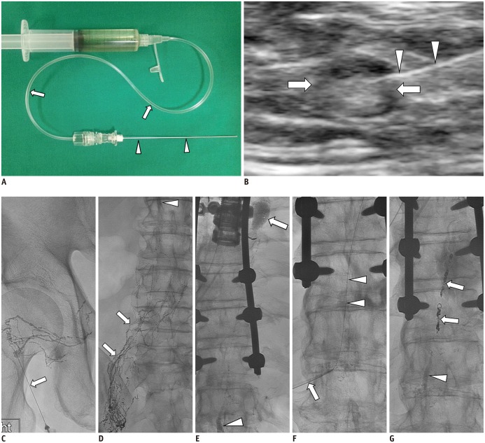
65-year-old man with history of esophageal cancer; status post total esophagectomy who presented with average of 2-3 L of left chylous effusion. A. 25-G spinal needle (arrowheads) is assembled to connecting tube (arrows) and is flushed with Lipiodol contained in syringe tube. B. Ultrasound-guided inguinal lymph node (arrows) access for initiation of intranodal LG using 25-G spinal needle (arrowheads). C. Initial spot image of right pelvis/inguinal region demonstrating needle (arrow) access of small lymph node and opacification of proximal lymphatic vessels and lymph node. D. Follow-up spot image of lower abdomen demonstrating upward move/flow of lymphatics (arrows) and appearance of cisterna chyli (arrowhead). E. Subsequent follow-up spot image of thoracolumbar region demonstrating continued upward flow of lymphatics with filling of cisterna chyli (arrowhead) and evidence of lymphatic leak showing in upper thoracic region with pooling of Lipiodol contrast (arrow). F. Cisterna chyli is percutaneously accessed using 22-G Chiba needle (arrow) and once it is accessed, microwire (arrowheads) is used to secure access by advancing it to mid thoracic duct. G. In this patient, microwire and microcatheter were unable to advance beyond leak along thoracic duct. Decision was made to coil embolize thoracic duct proximal to leak using several detachable coils (arrows) and it was further embolized using NBCA (arrowhead) down to cisterna chyli. On post-TDE day 1, chylous effusion completely stopped and chest tube drainage decreased to less than 50 mL from 3 L. LG = lymphangiography, NBCA = N-butyl-2-cyanoacrylate, TDE = thoracic duct embolization
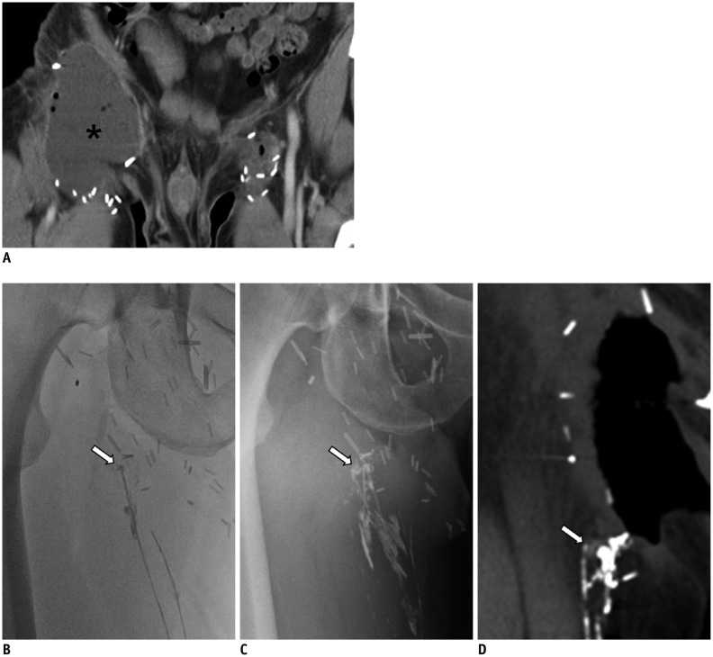
45-year-old man with right inguinal area swelling after deep inguinal lymph node dissection. A. Axial CT scan shows lymphocele (asterisk), which was subsequently dissected, in right inguinal area. Note minimal fluid collection at left superficial inguinal lymph node dissection site. B. Radiographic image obtained 30 minutes following Lipiodol injected into lymphatics of dorsum of foot, shows disruption of lymphatic with Lipiodol leakage (arrow) at entrance of open wound in right inguinal area. C. Radiographic image obtained 14 hours following Lipiodol injection, still shows leakage point (arrow). D. CT scan obtained 20 hours following Lipiodol injection shows disrupted lymphatic (arrow). Leakage was eliminated five days following LG. LG = lymphangiography
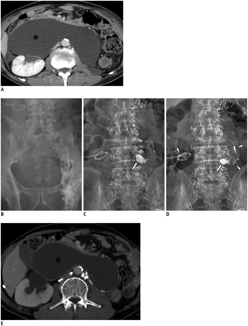
59-year-old man with history of nephroureterectomy and pelvic lymph node dissection. A. Axial CT scan shows large retroperitoneal lymphocele (asterisk) displacing both bowel and right kidney. B. Radiographic image obtained 20 minutes following Lipiodol injection into left inguinal lymph node shows good opacification of lymphatics in left pelvic cavity. C, D. Radiographic images obtained four and 16 hours following Lipiodol injection shows Lipiodol leakage (arrows) at 4th lumbar spine level and scattered Lipiodols (arrowheads) within lymphocele cavity as well as drainage tube. E. Axial CT scan obtained 20 days following initial LG shows decreased size of lymphocele (asterisk) and with decrease in amount of drainage. LG = lymphangiography
References
-
- Kortes N, Radeleff B, Sommer CM, Bellemann N, Ott K, Richter GM, et al. Therapeutic lymphangiography and CT-guided sclerotherapy for the treatment of refractory lymphatic leakage. J Vasc Interv Radiol. 2014;25:127–132. - PubMed
-
- Guermazi A, Brice P, Hennequin C, Sarfati E. Lymphography: an old technique retains its usefulness. Radiographics. 2003;23:1541–1558. discussion 1559-1560. - PubMed
-
- Matsumoto T, Yamagami T, Kato T, Hirota T, Yoshimatsu R, Masunami T, et al. The effectiveness of lymphangiography as a treatment method for various chyle leakages. Br J Radiol. 2009;82:286–290. - PubMed
-
- Kawasaki R, Sugimoto K, Fujii M, Miyamoto N, Okada T, Yamaguchi M, et al. Therapeutic effectiveness of diagnostic lymphangiography for refractory postoperative chylothorax and chylous ascites: correlation with radiologic findings and preceding medical treatment. AJR Am J Roentgenol. 2013;201:659–666. - PubMed
-
- Ngan H, Fok M, Wong J. The role of lymphography in chylothorax following thoracic surgery. Br J Radiol. 1988;61:1032–1036. - PubMed
Publication types
MeSH terms
LinkOut - more resources
Full Text Sources
Other Literature Sources
