

Adenocarcinoma of the sphenoid sinus
- PMID: 25469178
- PMCID: PMC4247863
- DOI: 10.11604/pamj.2014.18.284.4416
Adenocarcinoma of the sphenoid sinus
Abstract
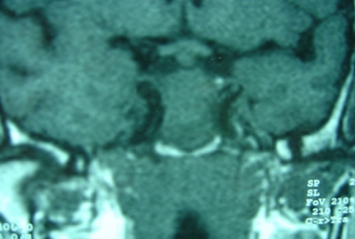
Adenocarcinomas of the sphenoid sinus are exceptional. In this paper, we report a new case with a review of the literature. Our patient was a 45-year-old man who presented with isolated retro orbital headache. CT and MRI suspected a malignat tumor of the sphenoid sinus. The patient underwent a debulking surgery. The final pathology carried out the diagnosis of primary adenocarcinoma. The patient died several months later from radiotherapy complications. Even if adenocarcinomas of the sphenoid sinus are exceptional, they should be considered in the differential diagnosis of sphenoid sinus masses. The prognosis is poor.
Keywords: Sphenoid sinus; adenocarcinoma; endonasal surgery; radiotherapy.
Figures


References
-
- Beaujeux R, Dietemann J, Brun F, Bourjat P. Scanographie et IRM des tumeurs et pseudo-tumeurs du sphénoïde. Feuillets de radiologie. 1994;34(3):214–229.
-
- DeMonte F, Ginsberg LE, Clayman GL. Primary malignant tumors of the sphenoidal sinus. Neurosurgery. 2000;46(5):1084–1092. - PubMed
-
- Grau C, Jakobsen MH, Harbo G, Svane-Knudsen V, Wedervang K, Larsen SK, Rytter C. Sino-nasal Cancer in Denmark 1982-1999: A Nationwide Survey. Acta Oncologica. 2001;40(1):19–23. - PubMed
-
- Cakmak O, Ergin TN, Aydin VM. Isolated sphenoid sinus adenocarcinoma: a case report. European archives of oto-rhino-laryngology. 2002;259(5):266–268. - PubMed
-
- Çolak A, Benli K, Dönmez T, Önol B. Papillary Carcinoma of the Sphenoid Sinus Associated with Sphenoid Sinus Abscess Presenting as Cavernous Sinus Syndrome: A Case Report. Journal of Neuro-Ophthalmology. 1990;10(1):18–20. - PubMed
Publication types
MeSH terms
LinkOut - more resources
Full Text Sources
Other Literature Sources
