Giant cell tumor of the patella: An uncommon cause of anterior knee pain
- PMID: 25469296
- PMCID: PMC4251109
- DOI: 10.3892/mco.2014.433
Giant cell tumor of the patella: An uncommon cause of anterior knee pain
Abstract
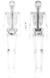
The patella is a rare site for the development of primary tumors. This is the case report of a giant cell tumor (GCT) occurring in the patella in a 25-year-old woman. The patient presented with a 1-year history of occasional right anterior knee pain. The radiological characteristics suggested a benign condition. The intraoperative pathological diagnosis was GCT of the bone. The lesion was treated by radical curettage with adjuvant therapy comprising phenol and ethanol and injection of calcium phosphate cement. Histologically, the tumor consisted of round or spindle-shaped mononuclear cells admixed with numerous osteoclastic giant cells. The patient was asymptomatic and there was no evidence of local recurrence or distant metastasis 16 months after surgery. Although rare, patellar GCT may be included in the differential diagnosis of anterior knee pain and/or swelling, particularly in young adults.
Keywords: anterior knee pain; giant cell tumor; imaging; patella.
Figures


References
-
- Singh J, James SL, Kroon HM, Woertler K, Anderson SE, Jundt G, Davies AM. Tumour and tumour-like lesions of the patella - a multicentre experience. Eur Radiol. 2009;19:701–712. - PubMed
-
- Casadei R, Kreshak J, Rinaldi R, Rimondi E, Bianchi G, Alberghini M, Ruggieri P, Vanel D. Imaging tumors of the patella. Eur J Radiol. 2013;82:2140–2148. - PubMed
-
- Athanasou NA, Bansal M, Forsyth R, Reid RP, Sapi Z. Giant cell tumor of bone. In: Fletcher CDM, Bridge JA, Hogendoorn PCW, Mertens F, editors. World Health Organization Classification of Tumours of Soft Tissue and Bone. 4th. IARC Press; Lyon: 2013. pp. 321–324.
-
- Chakarun CJ, Forrester DM, Gottsegen CJ, Patel DB, White EA, Matcuk GR., Jr Giant cell tumor of bone: review, mimics, and new developments in treatment. Radiographics. 2013;33:197–211. - PubMed
-
- Saglik Y, Yildiz Y, Basarir K, Tezen E, Guner D. Tumours and tumour-like lesions of the patella: a report of eight cases. Acta Orthop Belg. 2008;74:391–396. - PubMed
LinkOut - more resources
Full Text Sources
Other Literature Sources
