. 2015 Feb;27(2):160-74.
doi: 10.1111/nmo.12477. Epub 2014 Dec 3.

The Chicago Classification of esophageal motility disorders, v3.0

Affiliations

The Chicago Classification of esophageal motility disorders, v3.0

P J Kahrilas et al. Neurogastroenterol Motil. 2015 Feb.

Abstract

Background: The Chicago Classification (CC) of esophageal motility disorders, utilizing an algorithmic scheme to analyze clinical high-resolution manometry (HRM) studies, has gained acceptance worldwide.

Methods: This 2014 update, CC v3.0, developed by the International HRM Working Group, incorporated the extensive clinical experience and interval publications since the prior (2011) version.

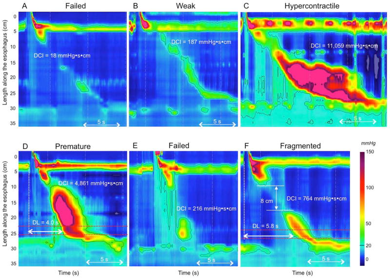
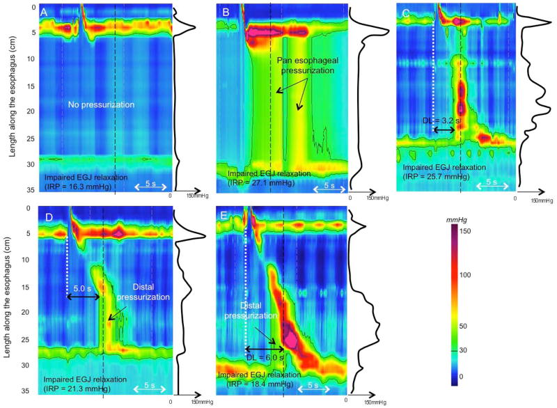
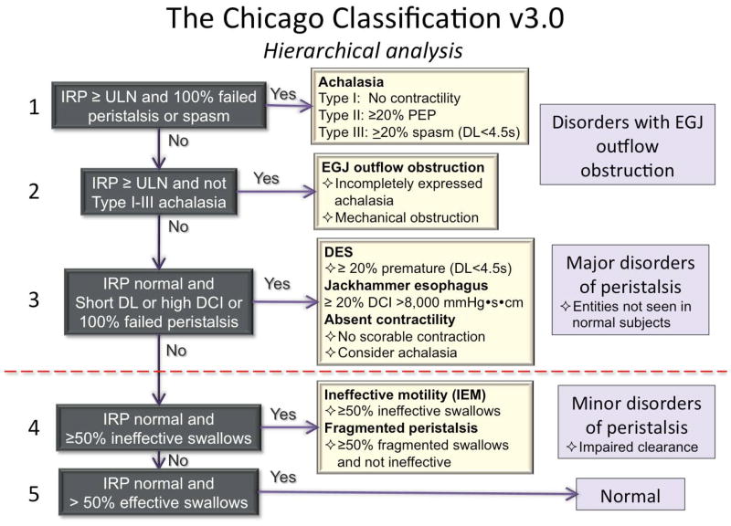
Key results: Chicago Classification v3.0 utilizes a hierarchical approach, sequentially prioritizing: (i) disorders of esophagogastric junction (EGJ) outflow (achalasia subtypes I-III and EGJ outflow obstruction), (ii) major disorders of peristalsis (absent contractility, distal esophageal spasm, hypercontractile esophagus), and (iii) minor disorders of peristalsis characterized by impaired bolus transit. EGJ morphology, characterized by the degree of overlap between the lower esophageal sphincter and the crural diaphragm and baseline EGJ contractility are also part of CC v3.0. Compared to the previous CC version, the key metrics of interpretation, the integrated relaxation pressure (IRP), the distal contractile integral (DCI), and the distal latency (DL) remain unchanged, albeit with much more emphasis on DCI for defining both hypo- and hypercontractility. New in CC v3.0 are: (i) the evaluation of the EGJ at rest defined in terms of morphology and contractility, (ii) 'fragmented' contractions (large breaks in the 20-mmHg isobaric contour), (iii) ineffective esophageal motility (IEM), and (iv) several minor adjustments in nomenclature and defining criteria. Absent in CC v3.0 are contractile front velocity and small breaks in the 20-mmHg isobaric contour as defining characteristics.

Conclusions & inferences: Chicago Classification v3.0 is an updated analysis scheme for clinical esophageal HRM recordings developed by the International HRM Working Group.

Keywords: achalasia; dysphagia; esophageal motility disorders; high-resolution manometry; ineffective esophageal motility.


Conflict of interest statement

COMPETING INTERESTS

Albert J Bredenoord: Given Imaging: research funding; Medical Measurement Systems: educational and research funding

Mark Fox: Given imaging: advisory board, consulting and educational; Sandhill Scientific: educational; Medical Measurement Systems: educational.

C Prakash Gyawali: Given imaging, educational

John E Pandolfino: Given imaging; consulting and educational

Serhat Bor: Medical Measurement Systems: educational and research funding

DO Castell: Sandhill Scientific Instruments; consultant and educational

Jeffrey L Conklin: Given imaging: consulting and educational

Sabine Roman: Given imaging: advisory board, consulting and educational

Phil Katz: Torax: consultant; Prizer Consumer: consultant

Jutta Keller: Given imaging: research funding, consulting and educational; Standard Imaging: consulting and educational

Taher Omari: Sandhill Scientific Instruments: consulting and research grant; Medical Measurement Systems: educational.

Edoardo Savarino: Given imaging; advisory board, consulting and educational

Felice Schnoll-Sussman: Given imaging; advisory board, consulting and educational

Daniel Sifrim: Given imaging; advisory board. Sandhill Scientific Instruments: research grant.

Rami Sweis: Given imaging; consulting and educational

Radu Tutuian: Sandhill Scientific Instruments: educational; Medical Measurement Systems: educational.

Marcelo F Vela: Given Imaging, Consulting

Frank Zerbib: Given imaging; advisory board, consulting, and educational.

Figures

Figure 1
Figure 1
The CDP represents the inflexion point in the contractile front propagation velocity in the distal esophagus. However, two inflexion points can sometimes be identified in instances of atypical peristaltic architecture (Panels A and B). Depending of the location of the inflexion point, DL measured from UES relaxation to the CDP might be either normal (>4.5 s) or reduced (upper location, Panel B). Adding the stipulation that the CDP must be localized to within 3 cm of the proximal margin of the LES (area between the 2 red dotted lines) prevents miscategorization in Panels A and B. In instances of compartmentalized pressurization (Panel C) the CDP is localized along a pressure above the intra-bolus pressure (50 mmHg isobaric contour on Panel C), not to the moment of compartmentalized pressurization.
Figure 2
Figure 2
Lack of relevance of CFV in the identification of DES. The CFV can be elevated (>9 cm/s) with a reduced DL (<4.5 s) (Panel A, a patient with symptoms of dysphagia and chest pain). However, in cases with a normal DL, an elevated CFV might be seen as the consequence of atypical contraction architecture (patients without dysphagia in Panels B and C) or a large break in the contractile front (Panel D, patient with IEM and a negative CFV value on this swallow).
Figure 3
Figure 3
Normal esophageal contraction followed by hyperconctractility of the LES (Panel A). Including the LES (the 4th contractile segment defined by Clouse) in the DCI measurement (white dashed box) result in a diagnosis of hypercontractility. In the case of borderline DCI (Panel B) including LES in the DCI measurement also results in the diagnosis of hypercontractility.
Figure 4
Figure 4
sophagogastric junction (EGJ) morphology subtypes. For each panel the instantaneous spatial pressure variation plot corresponding to the red line on the pressure topography plot is illustrated by the black line to the right. The two main EGJ components are the LES and CD, which cannot be independently quantified when they are superimposed as with a type I EGJ (Panel A). The respiratory inversion point (RIP), shown by the white horizontal dashed line, lies near the proximal margin of the EGJ. During inspiration (I) EGJ pressure increases, whereas it decreases during expiration (E). Type II EGJ pressure morphology is illustrated in Panel B. Note the 2 peaks on the instantaneous spatial pressure variation plot; the nadir pressure between the peaks is greater than the intra gastric pressure. The RIP is at the level of the CD. Panels C and D correspond to type III EGJ pressure morphology defined as the presence of 2 peaks of the instantaneous spatial pressure variation plot with the nadir pressure between the peaks equal to or less than intragastric pressure. The RIP is at the CD with type IIIa (Panel C) whereas it is at the level of the LES in IIIb (Panel D).
Figure 5
Figure 5
Contractile vigor and contraction pattern. Contractile vigor is assessed using the DCI: a contraction with a DCI <100 mmHg·s·cm is failed (Panel A); a contraction with a DCI >100 but <450 mmHg·s·cm is weak (Panel B); and a hypercontractile swallow is defined as a DCI >8,000 mmHg·s·cm (Panel C). Premature contraction is defined as a distal latency (DL) <4.5 s (Panel D). A weak contraction (DCI <450 mmHg·s·cm) with a reduced distal latency is considered failed (Panel E). A contraction with a normal DCI (450–8,000 mmHg·s·cm) and a break >5 cm is a fragmented contraction (Panel F).
Figure 6
Figure 6
Disorders associated with EGJ outflow obstruction. Impaired EGJ relaxation is evident by an IRP >15 mmHg. Instantaneous pressure along the black dashed line is represented by the black line on the right side of each panel. In type I achalasia, there is no esophageal contraction and no esophageal pressurization (Panel A). Type II achalasia is characterized by panesophageal pressurization and absence of a peristaltic contraction (Panel B). Note that pressurization corresponds to homogeneous pressure along the spatial pressure variation plot. In type III achalasia, there are at least 20% premature contractions, defined as DL <4.5 s. (Panel C). Note the multiple peaks corresponding to contractions along the spatial pressure variation plot. EGJ outflow obstruction may represent achalasia (Panel D); it might also be the consequence of a mechanical obstruction (Panel E) such as a distal esophageal stenosis.
Figure 7
Figure 7
Hierarchical algorithm for the interpretation of HRM studies with CC v3.0.

References

    1. Gyawali CP. High resolution manometry: the Ray Clouse legacy. Neurogastroenterol Motil. 2012 Mar 24;(Suppl 1):2–4. - PubMed
    1. Pandolfino JE, Fox MR, Bredenoord AJ, Kahrilas PJ. High-resolution manometry in clinical practice: utilizing pressure topography to classify oesophageal motility abnormalities. Neurogastroenterol Motil. 2009;21:796–806. - PMC - PubMed
    1. Bredenoord AJ, Fox M, Kahrilas PJ, et al. Chicago classification criteria of esophageal motility disorders defined in high resolution esophageal esophageal pressure topography (EPT) Neurogastroenterol Motil. 2012;24 (Suppl 1):57–65. - PMC - PubMed
    1. Bogte A, Bredenoord AJ, Oors J, Siersema PD, Smout AJ. Normal values for esophageal high-resolution manometry. Neurogastroenterol Motil. 2013;25:762–e579. - PubMed
    1. Shi Y, Xiao Y, Peng S, Lin J, Xiong L, Chen M. Normative data of high-resolution impedance manometry in the Chinese population. J Gastroenterol Hepatol. 2013;28:1611–1615. - PubMed

Publication types

MeSH terms