. 2014 Dec 3;9(12):e113799.
doi: 10.1371/journal.pone.0113799. eCollection 2014.

HMGB1 promotes a p38MAPK associated non-infectious inflammatory response pathway in human fetal membranes

Affiliations

HMGB1 promotes a p38MAPK associated non-infectious inflammatory response pathway in human fetal membranes

Sarah Bredeson et al. PLoS One. .

Abstract

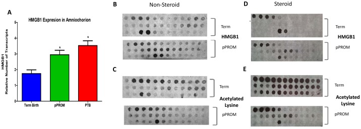
Objective: Spontaneous preterm birth (PTB) and preterm prelabor rupture of membranes (pPROM) are major pregnancy complications often associated with a fetal inflammatory response. Biomolecular markers of this fetal inflammatory response to both infectious and non-infectious risk factors and their contribution to PTB and pPROM mechanism are still unclear. This study examined fetal membrane production, activation and mechanistic properties of high mobility group box 1 (HMGB1) as a contributor of the non-infectious fetal inflammatory response.

Materials and methods: HMGB1 transcripts and active HMGB1 were profiled in fetal membranes and amniotic fluids collected from PTB and normal term birth. In vitro, normal term not in labor fetal membranes were exposed to lipopolysaccharide (LPS) and water soluble cigarette smoke extract (CSE). HMGB1-transcripts and its protein concentrations were documented by RT-PCR and ELISA. Recombinant HMGB1 treated membranes and media were subjected to RT-PCR for HMGB1 receptors, mitogen activated protein kinase pathway analysis, cytokine levels, and Western blot for p38MAPK.

Results: HMGB1 expression and its active forms were higher in PTB and pPROM than normal term membranes and amniotic fluid samples. Both LPS and CSE enhanced HMGB1 expression and release in vitro. Fetal membrane exposure to HMGB1 resulted in increased expression of TLR2 and 4 and dose-dependent activation of p38MAPK-mediated inflammation.

Conclusions: HMGB1 increase by fetal membrane cells in response to either oxidative stress or infection can provide a positive feedback loop generating non-infectious inflammatory activation. Activation of p38MAPK by HMGB1 promotes development of the senescence phenotype and senescence associated sterile inflammation. HMGB1 activity is an important regulator of the fetal inflammatory response regardless of infection.


Conflict of interest statement

Competing Interests: The authors have declared that no competing interests exist.

Figures

Figure 1
Figure 1. HMGB1 Expression in Fetal Membrane and Amniotic Fluid Cases and Controls A.
Bar graph shows HMGB1 expression in fetal membranes as the mean and standard error of relative number of transcripts as determined by real time PCR. GAPDH was used as an internal control. Comparison among the clinical samples from women at term not in labor (Term Birth), with preterm premature rupture of membranes (pPROM), and with preterm birth with intact membranes (PTB). (ANOVA, *p<0.05). B-E. HMGB1 and acetylated lysine in human amniotic fluid (AF). B. pPROM AF without steroids has a greater amount of HMGB1 than term AF. C. Term AF has a greater amount of total protein acetylation than pPROM AF without steroids. D. pPROM AF with glucocorticoids has a greater amount of HMGB1 than term AF. E. Term AF has a greater amount of total protein acetylation than pPROM AF with steroids. However, HMGB1 retains acetylation in pPROM despite deacetylation of proteins by administered steroids (D vs. E) as seen by the preserved HMGB1 pattern. Interestingly, more proteins are acetylated in pPROM AF without steroid use than with steroid administration (C vs. E). In both term and pPROM, HMGB1 shows a consistent expression pattern (B vs. D).
Figure 2
Figure 2. HMGB1, Receptor, and Proinflammatory Cytokine Expression in Fetal Membranes.
Bar graphs show A. HMGB1 expression as the mean and standard error of relative number of transcripts as determined by real time PCR. GAPDH was used as an internal control. Comparison among control, cigarette smoke extract-stimulated (CSE), and lipopolysaccharide-stimulated (LPS) fetal membranes at term. B. HMGB1 concentration as the mean and standard error of relative intensity as determined by competitive enzyme immunoassay ELISA. Note: Intensity of color detected is inversely proportional to the HMGB1 concentration. Comparison among control, cigarette smoke extract-stimulated (CSE), and lipopolysaccharide-stimulated (LPS) culture media at term. C–E. HMGB1 receptor expression in HMGB1-stimulated (1, 5, 10, 50 ng/ml) fetal membranes as the mean and standard error of relative number of transcripts as determined by real time PCR. GAPDH was used as an internal control. C. Comparison of RAGE expression among HMGB1 treated fetal membrane samples. D. Comparison of TLR2 expression among HMGB1 treated fetal membrane samples. E. Comparison of TLR4 expression among HMGB1 treated fetal membrane samples. F–H. Cytokine concentration in HMGB1-stimulated (1, 5, 10, 50 ng/ml) fetal membrane culture media as the mean and standard error of relative concentration as determined by multiplex human cytokine panel analysis. F. Comparison of IL-1β concentration among HMGB1 treated culture media samples. G. Comparison of TNFα concentration among HMGB1 treated culture media samples. H. Comparison of IL-6 concentration among HMGB1 treated culture media samples. (ANOVA, *p<0.05).
Figure 3
Figure 3. Pp38 MAPK and NF-κB Pathways Activation by HMGB1.
A–B. Bar graphs show MAP kinase protein concentration in HMGB1-stimulated (1, 5, 10, 50 ng/mL) fetal membranes as the mean and standard error of relative intensity as determined by multiplex human MAPK protein panel analysis. A. Comparison of phosphorylated-p38 (Pp38) concentration among HMGB1-treated fetal membrane samples. B. Comparison of HSP27 concentration among HMGB1-treated fetal membrane samples. C. Dose dependent activation of Pp38MAPK after HMGB1 treatment where maximum activation was seen after 50 ng/ml HMGB1 treatment. D–E. Bar graphs show cytokine concentrations in HMGB1 (50 ng/ml) and HMGB1+SB 203580 (30 uM) p38MAPK inhibitor treated fetal membrane culture media. D. Comparison of IL-6 concentration among control, SB 203580 alone, HMGB1alone, and HMGB1+SB203580 cultures. E. Comparison of IL-8 concentration among control, SB 203580 alone, HMGB1alone, and HMGB1+SB203580 cultures. F. p38MAPK dependent NF-κB activation. HMGB1 (50 ng/ml) increased RelA phosphorylation. (ANOVA, *p<0.05).
Figure 4
Figure 4. SA-β-Gal staining.
A–B. Pp38 MAPK mediated senescence phenotype (SP) development was determined using Senescence Associated β -Galactosidase (SA β-Gal) assay. SP was seen as blue staining cells and the number of these cells were higher after 50 ng/ml of HMGB1 treatment (B) compared to control (A). C. Bar graph on the right shows percentage of SA β-Gal staining cells after 50 ng/ml of HMGB1 treatment compared to untreated control.
Figure 5
Figure 5. HMGB1 SASP Cytokine Production.
Bar graphs show cytokine concentrations in p38MAPK inhibitor SB203580, HMGB1 inhibitor (GA), and HMGB1treated, and simultaneous HMGB1 with SB203580 (30 uM), GA (100 uM), or a combination of both SB203580 and GA treated fetal membrane culture media. A. Comparison of IL-6 concentration among control, SB 203580 alone, GA alone, HMGB1alone, HMGB1+SB203580, HMGB1+GA, and HMGB1+SB+GA cultures. B. Comparison of IL-8 concentration among control, SB 203580 alone, GA alone, HMGB1alone, HMGB1+SB203580, HMGB1+GA, and HMGB1+SB+GA cultures. (ANOVA; *p <0.05).
Figure 6
Figure 6. Model of HMGB1 Mediated Inflammatory Response.
Proposed mechanistic pathway of HMGB1-mediated signaling in human fetal cells. In Phase 1, infectious or non-infectious risk factors associated with spontaneous preterm birth and preterm premature rupture of the membranes cause reactive oxygen species (ROS) formation from mitochondrial and non-mitochondrial origins in fetal membrane cells (44). This ROS causes nuclear HMGB1 to translocate into the cytoplasm. In the cytoplasm, HMGB1 is acetylated or modified by lysosomes and secreted either due to cellular injury (necrosis) or through carriers. HMGB1 can also be secreted in the non-acetylated form. In Phase 2, a feed-forward loop can be established by HMGB1 to enhance the inflammatory condition of the fetal cells. Depending on the concentration, HMGB1 mediates an inflammatory condition to neighboring cells. Higher concentrations of HMGB1 induce TLR2 and lower concentrations promote TLR4-mediated signaling resulting in p38MAPK activation mediated through activation of MAPK series of signaling molecules (MAPKKK →MAPKK →MAPK) (45). p38MAPK can lead to development of senescence phenotype (SP) in human fetal cells. Senescing cells cause a unique inflammatory signature that is known as “Senescence Associated Secretory Phenotype (SASP)”. With SASP, a unique set of proinflammatory cytokines, chemokines, growth factors, angiogenic factors and matrix degrading enzymes and their inhibitors may be activated independently through the p38MAPK pathway or with possible involvement of the NF-κB pathway (34; 35). In the absence of intraamniotic infection this sterile inflammation may produce similar outcomes as the well-defined infection-associated NF-κB pathway. Furthermore, with or without intraamniotic infection, this interaction may also activate HMGB1 and the autocrine effect of HMGB1 will continue the vicious cycle of inflammatory events until delivery. Inflammation seen at term, and in cases with PTB and pPROM, may arise from a senescence-oriented pathway mediated through generation of HMGB1 by oxidative stress or tissue injury.

References

    1. Combs CA, Gravett M, Garite TJ, Hickok DE, Lapidus J, et al. (2014) Amniotic fluid infection, inflammation, and colonization in preterm labor with intact membranes. Am J Obstet Gynecol 210:125. - PubMed
    1. Romero R, Espinoza J, Goncalves LF, Kusanovic JP, Friel L, et al. (2007) The role of inflammation and infection in preterm birth. Semin Reprod Med 25:21–39. - PMC - PubMed
    1. Romero R, Chaiworapongsa T, Alpay SZ, Xu Y, Hussein Y, et al. (2011) Damage-associated molecular patterns (DAMPs) in preterm labor with intact membranes and preterm PROM: a study of the alarmin HMGB1. J Matern Fetal Neonatal Med 24:1444–1455. - PMC - PubMed
    1. Menon R, Fortunato SJ (2009) Distinct pathophysiologic pathways induced by in vitro infection and cigarette smoke in normal human fetal membranes. Am J Obstet Gynecol 200:334–338. - PubMed
    1. Bartling B, Fuchs C, Somoza V, Niemann B, Silber RE, et al. (2007) Lung level of HMBG1 is elevated in response to advanced glycation end product-enriched food in vivo. Mol Nutr Food Res 51:479–487. - PubMed

Publication types

MeSH terms

LinkOut - more resources