. 2014 Dec 3;9(12):e114472.
doi: 10.1371/journal.pone.0114472. eCollection 2014.

CX3CR1 signaling on monocytes is dispensable after intracerebral hemorrhage

Affiliations

CX3CR1 signaling on monocytes is dispensable after intracerebral hemorrhage

Roslyn A Taylor et al. PLoS One. .

Abstract

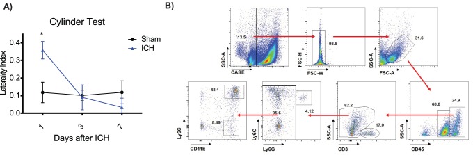
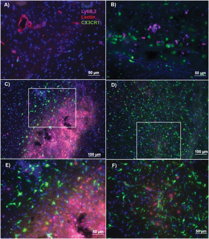
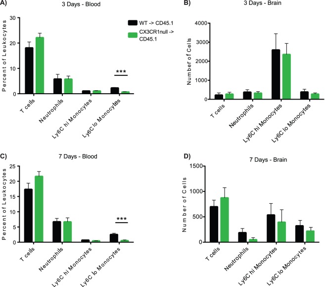
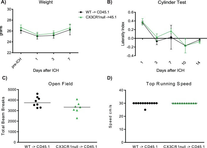
Intracerebral hemorrhage is a subset of stroke for which there is no specific treatment. The Ly6Chi CCR2+ monocytes have been shown to contribute to acute injury after intracerebral hemorrhage. The other murine monocyte subset expresses CX3CR1 and lower Ly6C levels, and contributes to repair in other disease models. We hypothesized that the Ly6Clo CX3CR1+ monocytes would contribute to recovery after intracerebral hemorrhage. Intracerebral hemorrhage was modeled by blood injection in WT and CX3CR1-null bone marrow chimeras. Neurological outcomes and leukocyte recruitment were quantified at various time points. Functional outcomes were equal at 1, 3, 7, and 14 days after intracerebral hemorrhage in both genotypes. No differences were observed in leukocyte recruitment between genotypes on either 3 or 7 days after intracerebral hemorrhage. A few hundred Ly6Clo monocytes were found in the ipsilateral hemisphere in each genotype and they did not change over time. Peripherally derived CX3CR1+ monocytes were observed in the perihematomal brain 7 and 14 days after intracerebral hemorrhage. Our data suggests CX3CR1 signaling on monocytes does not play an influential role in acute injury or functional recovery after intracerebral hemorrhage and therefore CX3CR1 is not a therapeutic target to improve outcome after intracerebral hemorrhage.


Conflict of interest statement

Competing Interests: The authors have declared that no competing interests exist.

Figures

Figure 1
Figure 1. Ly6Clo monocytes are present in the ipsilateral hemisphere 72 hours after ICH.
A) WT mice were cylinder tested 1, 3, and 7 days after either sham or ICH surgery. Means graphed with s.e.m., n = 6–8, *p<0.05 B) Ly6Clo monocytes are found in the ipsilateral hemisphere 3 days after ICH when mice recover by cylinder test. Cells were gated on live/dead, singlets, leukocytes were found by forward and side scatter, and then further gated to isolate populations as shown in these representative plots.
Figure 2
Figure 2. CX3CR1+, Ly6Clo monocytes are present in perihematomal brain in CX3CR1-null BM chimeras.
Mice were injected intracardially with 150 ug tomato lectin to delineate vasculature. Brain slices were stained with Ly6B.2 antibody to differentiate Ly6Chi monocytes and neutrophils from Ly6Clo CX3CR1+ monocytes and Dapi nuclear stain. A) Intracardiac injection of lectin stains vasculature in the brain– 20x. B) At 72 hours after ICH, distinct subsets of amoeboid Ly6B.2+ (Ly6Chi monocytes or neutrophils) and GFP+ (CX3CR1+) monocytes are seen in the perihematomal region. 20x. C) At 7 days after ICH, amoeboid, Ly6B.2,CX3CR1+ cells are seen in the brain parenchyma surrounding the ICH cavity. The center of the ICH cavity is filled with Ly6B.2+ (Ly6Chi) monocytes or neutrophils. Tomato lectin staining is also seen in the center of the ICH cavity, most likely due to blood brain barrier breakdown at this time point leading to nonspecific lectin staining of myeloid cells. 10x. White box indicates the inset shown in panel E. D) At 14 days, amoeboid, CX3CR1+ cells can still be seen in the perihematomal region, however more CX3CR1+ cells have a ramified morphology. No Ly6B.2+ (Ly6Chi monocytes or neutrophils) cells are found. Overall blood-brain barrier disruption is improved leading to less lectin staining in the cavity, although there remain some lectin+ cells (CX3CR1+ monocytes and CX3CR1 microglia). 10x. White box indicates inset shown in panel F. E) 20x image of panel C – perihematomal region 7 days after ICH F) 20x image of panel D – perihematomal region 14 days after ICH. Blue – Dapi, GFP – CX3CR1, Red – Tomato lectin, Pink – Ly6B.2, n = 5.
Figure 3
Figure 3. CX3CR1-deficiency on Ly6Clo monocytes does not affect leukocyte recruitment to the ipsilateral hemisphere.
A) Percent of leukocytes in the blood 3 days after ICH. CX3CR1 BM chimeras have a reduction in percentage of Ly6Clo CX3CR1+ monocytes. B) At 3 days after ICH, WT BM chimeras and CX3CR1-null BM chimeras have equal numbers of T cells, Ly6Chi monocytes, Ly6Clo CX3CR1+ monocytes, and neutrophils in the ipsilateral hemisphere. The Ly6Chi monocytes constitute the most numerous cell population recruited to the brain at 3 days in both genotypes. T cells, neutrophils, and Ly6Clo CX3CR1+ monocytes are recruited in roughly equal numbers. n = 10 C) Percent of leukocytes in the blood 7 days after ICH. Similar to 3 days, the CX3CR1 BM chimeras have a smaller percentage Ly6Clo CX3CR1+ monocytes in the blood. D) At 7 days, the numbers of T cells have increased, Ly6Chi monocytes and neutrophils decreased, with no change in the numbers of Ly6Clo CX3CR1+ monocytes. Like at 3 days, peripheral leukocyte populations do not differ between WT BM chimeras and CX3CR1-null chimeras. Bars indicate mean ± s.e.m. n = 9.
Figure 4
Figure 4. CX3CR1 deficiency on monocytes does not affect functional outcomes days 1–14 after ICH.
A) WT BM chimeras and CX3CR1-null BM chimeras have similar weight loss and recovery. Means graphed with s.e.m.,n = 8–21, p>0.05 at all time points B) WT BM chimeras and CX3CR1-null BM chimeras have equal left forelimb weakness 24 hours after ICH and recover equally over the first 14 days by cylinder test. Means graphed with s.e.m., n = 8, p>0.05 at all time points C) Open field test shows CX3CR1 deficiency on monocytes does not impact spontaneous locomotor activity at 7 or 14 days after ICH. Line represents mean number of total beam breaks. n = 7–8, p>0.05 at each time point. D) CX3CR1-null BM chimeras perform equally well on the forced run test 7 and 14 days after ICH. Line represents median speed. n = 8–12, p>0.05 at each time point.

References

    1. Manno E (2012) Update on intracerebral hemorrhage. Continuum (Minneapolis, Minn) 18:598–610. - PubMed
    1. Qureshi A, Mendelow A, Hanley D (2009) Intracerebral haemorrhage. Lancet 373:1632–1644. - PMC - PubMed
    1. Keep R, Hua Y, Xi G (2012) Intracerebral haemorrhage: mechanisms of injury and therapeutic targets. Lancet neurology 11:720–731. - PMC - PubMed
    1. Hammond M, Ai Y, Sansing L (2012) Gr1+ Macrophages and Dendritic Cells Dominate the Inflammatory Infiltrate 12 Hours After Experimental Intracerebral Hemorrhage. Translational stroke research 3. - PMC - PubMed
    1. Hammond MD, Taylor RA, Mullen MT, Ai Y, Aguila HL, et al. (2014) CCR2+Ly6Chi Inflammatory Monocyte Recruitment Exacerbates Acute Disability Following Intracerebral Hemorrhage. J Neurosci 34:3901–3909. - PMC - PubMed

Publication types

LinkOut - more resources