. 2014 Dec 3;9(12):e114249.
doi: 10.1371/journal.pone.0114249. eCollection 2014.

Lenalidomide induces lipid raft assembly to enhance erythropoietin receptor signaling in myelodysplastic syndrome progenitors

Affiliations

Lenalidomide induces lipid raft assembly to enhance erythropoietin receptor signaling in myelodysplastic syndrome progenitors

Kathy L McGraw et al. PLoS One. .

Abstract

Anemia remains the principal management challenge for patients with lower risk Myelodysplastic Syndromes (MDS). Despite appropriate cytokine production and cellular receptor display, erythropoietin receptor (EpoR) signaling is impaired. We reported that EpoR signaling is dependent upon receptor localization within lipid raft microdomains, and that disruption of raft integrity abolishes signaling capacity. Here, we show that MDS erythroid progenitors display markedly diminished raft assembly and smaller raft aggregates compared to normal controls (p = 0.005, raft number; p = 0.023, raft size). Because lenalidomide triggers raft coalescence in T-lymphocytes promoting immune synapse formation, we assessed effects of lenalidomide on raft assembly in MDS erythroid precursors and UT7 cells. Lenalidomide treatment rapidly induced lipid raft formation accompanied by EpoR recruitment into raft fractions together with STAT5, JAK2, and Lyn kinase. The JAK2 phosphatase, CD45, a key negative regulator of EpoR signaling, was displaced from raft fractions. Lenalidomide treatment prior to Epo stimulation enhanced both JAK2 and STAT5 phosphorylation in UT7 and primary MDS erythroid progenitors, accompanied by increased STAT5 DNA binding in UT7 cells, and increased erythroid colony forming capacity in both UT7 and primary cells. Raft induction was associated with F-actin polymerization, which was blocked by Rho kinase inhibition. These data indicate that deficient raft integrity impairs EpoR signaling, and provides a novel strategy to enhance EpoR signal fidelity in non-del(5q) MDS.


Conflict of interest statement

Competing Interests: KM has read the journal's policy and the authors of this manuscript have the following competing interests: AFL was a consultant for Celgene. This does not alter the authors' adherence to PLOS ONE policies on sharing data and materials.

Figures

Figure 1
Figure 1. MDS erythroid precursors are deficient in membrane lipid rafts.
(A) Representative micrographs of primary erythroid precursors from an MDS patient (top) compared to normal donor (bottom) erythroid progenitors identified by confocal immunofluorescence microscopy. Primitive erythroids express CD71+ (green) and cKit+ (purple); lipid rafts display red fluorescence, DAPI (blue), merged image is shown in the last panel. Bars represent 25 µm. Enlarged images illustrate lipid rafts at 630x with 6-zoom magnification. Bars represent 5 µm. (B) Statistically significant decrease in the number of rafts per cell in MDS erythroid precursors compared to normal donor erythroid cells, graph represents mean ± SE. (C) Statistically significant decrease in the size of raft aggregates in primary erythroids compared to normal erythroids. Mean ± SE.
Figure 2
Figure 2. Lenalidomide induces raft aggregation in UT7 cells and MDS erythroid progenitors.
(A) Dot blot detection of GM-1 membrane fractionation in UT7 cells treated with 1 U/ml Epo or 1 µM LEN for 1 hr. Rafts are located predominantly in fraction 2 (all raft fractions 1–3) whereas non-raft fractions are represented by fractions 4–6. Numbers represent densitometry analysis of combined raft fractions 1–3. (B) Immunofluorescence of UT7 cells, DAPI (blue), rafts (red), and merged image showing marked accumulation of rafts after lenalidomide treatment. Bars represent 20 µm. (C) Representative micrograph of MDS primary erythroid precursors identified by CD71+ (green) and cKit+ (purple) expression, untreated (top) and lenalidomide treated (bottom) showing increased raft aggregation following drug treatment. Bars represent 25 µm. Enlarged image from inset shown. (D) There is a statistically significant increase in the size of raft aggregates of MDS erythroid cells that is not observed in normal erythroid cells. (E) Comparison of lipid raft size in lenalidomide responding MDS patient erythroid precursors compared to non-responders.
Figure 3
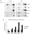
Figure 3. Lenalidomide induces recruitment of EpoR and signaling effectors into lipid rafts.
(A) Western blot of cell fractions showing lipid rafts primarily in fraction 2 and non-raft fractions in 5 and 6. Lenalidomide induces recruitment of EpoR, JAK2, and STAT5 into raft fractions while displacing CD45. Lyn kinase serves as an additional marker for lipid raft fractionation, and is also increased in raft fractions after lenalidomide treatment. (B) Relative raft protein expression by densitometry analysis.
Figure 4
Figure 4. Lenalidomide enhances EpoR signaling and erythroid colony growth.
(A) Western blot showing increased and prolonged JAK2 and STAT5 phosphorylation in UT7 cells treated with 1 U/ml rhEpo and 1 µM lenalidomide pretreatment for 1 h. (B) STAT5 EMSA showing increased and sustained binding of STAT5 to DNA in UT7 cells pretreated with lenalidomide. K562 cells were used as controls. Numbers represent densitometry analysis of lenalidomide + rhu-Epo treatment compared to Epo treatment alone at each time point. (C) Flow cytometry histograms showing an increase in P-STAT5 in primary erythroid progenitors identified as CD45dim, CD71high, and GlyAlow with MFI of the 95th% provided. (D) Colony forming capacity of UT7 cells pretreated with or without lenalidomide. 5 U/ml of rhEpo was added to the indicated wells with each performed in triplicate. Colonies were counted after 7 d incubation and are represented as mean ± SE. (E) Colony forming capacity of MDS BM-MNC showing more than a 2-fold increase of BFU-E after lenalidomide pretreatment without a marked increase in mixed lineage CFU-GEMM.
Figure 5
Figure 5. ROCK inhibition abrogates lenalidomide induced accumulation of lipid rafts.
(A) Dot blot detection of GM-1 in UT7 cells treated with lenalidomide with or without the ROCK inhibitor, Y-27632 (ROCKi), pretreatment showing a decrease in raft fractionation with combination treatment. Rafts are located in fractions 1 and 2, while non-raft fractions are 4–6. Numbers represent densitometry analysis of combined raft fractions 1 and 2. (B) Western blot of Lyn kinase indicative of raft fractionation (fractions 1 and 2) showing an increase after lenalidomide treatment that is partially blocked with ROCKi pretreatment. Numbers represent densitometry analysis of combined raft fractions 1–3. (C) Immunofluorescence of rafts (red), DAPI (blue), and merged image showing inhibition of lenalidomide induced raft formation with ROCKi pretreatment. Bars represent 20 µm.
Figure 6
Figure 6. Lenalidomide induces actin polymerization that is blocked by ROCK inhibition.
(A) Confocal microscopy at 630x and (B) 630x with 4x zoom of phalloidin (green) used to detect actin polymerization, DAPI (blue), and merged image. Lenalidomide treatment induced actin polymerization that was inhibited by pre-treatment with ROCKi, Y-27632. Bars represent 20 µm (A) and 10 µm (B).

References

    1. Hoefsloot LH, van Amelsvoort MP, Broeders LC, van der Plas DC, van Lom K, et al. (1997) Erythropoietin-induced activation of STAT5 is impaired in the myelodysplastic syndrome. Blood 89:1690–1700. - PubMed
    1. Ebert BL, Galili N, Tamayo P, Bosco J, Mak R, et al. (2008) An erythroid differentiation signature predicts response to lenalidomide in myelodysplastic syndrome. PLoS medicine 5:e35. - PMC - PubMed
    1. Fuhler GM, Blom NR, Coffer PJ, Drayer AL, Vellenga E (2007) The reduced GM-CSF priming of ROS production in granulocytes from patients with myelodysplasia is associated with an impaired lipid raft formation. Journal of leukocyte biology 81:449–457. - PubMed
    1. McGraw KL, Fuhler GM, Johnson JO, Clark JA, Caceres GC, et al. (2012) Erythropoietin receptor signaling is membrane raft dependent. PloS one 7:e34477. - PMC - PubMed
    1. Xavier R, Brennan T, Li Q, McCormack C, Seed B (1998) Membrane compartmentation is required for efficient T cell activation. Immunity 8:723–732. - PubMed

Publication types

MeSH terms