

The spatial and signal characteristics of physiologic high frequency oscillations
- PMID: 25470216
- PMCID: PMC5123742
- DOI: 10.1111/epi.12851
The spatial and signal characteristics of physiologic high frequency oscillations
Abstract
Objectives: To study the incidence, spatial distribution, and signal characteristics of high frequency oscillations (HFOs) outside the epileptic network.
Methods: We included patients who underwent invasive evaluations at Yale Comprehensive Epilepsy Center from 2012 to 2013, had all major lobes sampled, and had localizable seizure onsets. Segments of non-rapid eye movement (NREM) sleep prior to the first seizure were analyzed. We implemented a semiautomated process to analyze oscillations with peak frequencies >80 Hz (ripples 80-250 Hz; fast ripples 250-500 Hz). A contact location was considered epileptic if it exhibited epileptiform discharges during the intracranial evaluation or was involved ictally within 5 s of seizure onset; otherwise it was considered nonepileptic.
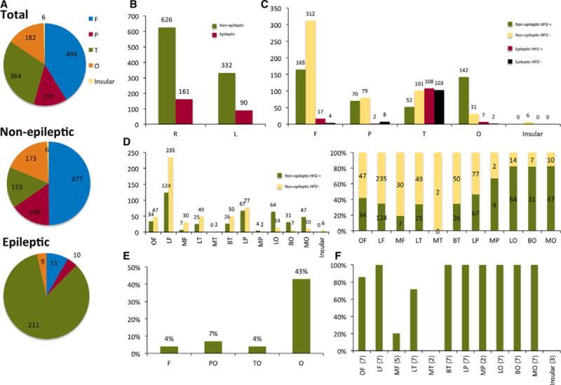
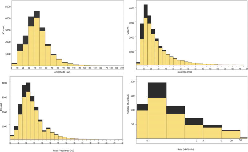
Results: We analyzed recordings from 1,209 electrode contacts in seven patients. The nonepileptic contacts constituted 79.1% of the total number of contacts. Ripples constituted 99% of total detections. Eighty-two percent of all HFOs were seen in 45.2% of the nonepileptic contacts (82.1%, 47%, 34.6%, and 34% of the occipital, parietal, frontal, and temporal nonepileptic contacts, respectively). The following sublobes exhibited physiologic HFOs in all patients: Perirolandic, basal temporal, and occipital subregions. The ripples from nonepileptic sites had longer duration, higher amplitude, and lower peak frequency than ripples from epileptic sites. A high HFO rate (>1/min) was seen in 110 nonepileptic contacts, of which 68.2% were occipital. Fast ripples were less common, seen in nonepileptic parietooccipital regions only in two patients and in the epileptic mesial temporal structures.
Conclusions: There is consistent occurrence of physiologic HFOs over vast areas of the neocortex outside the epileptic network. HFOs from nonepileptic regions were seen in the occipital lobes and in the perirolandic region in all patients. Although duration of ripples and peak frequency of HFOs are the most effective measures in distinguishing pathologic from physiologic events, there was significant overlap between the two groups.
Keywords: Electrocorticography; Epilepsy; High frequency oscillations; Intracranial EEG; Ripples.
Wiley Periodicals, Inc. © 2014 International League Against Epilepsy.
Figures
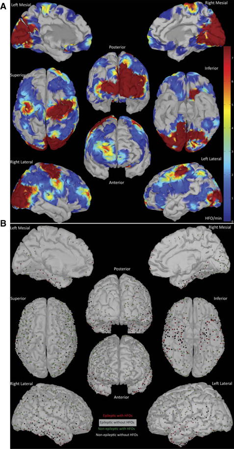
 , A contact with HFOs in an epileptic site;
 , a contact without HFOs in an epileptic site;
 , a contact with HFOs in a nonepileptic site; ○,a contact without HFOs in a nonepileptic site. Epilepsia © ILAE
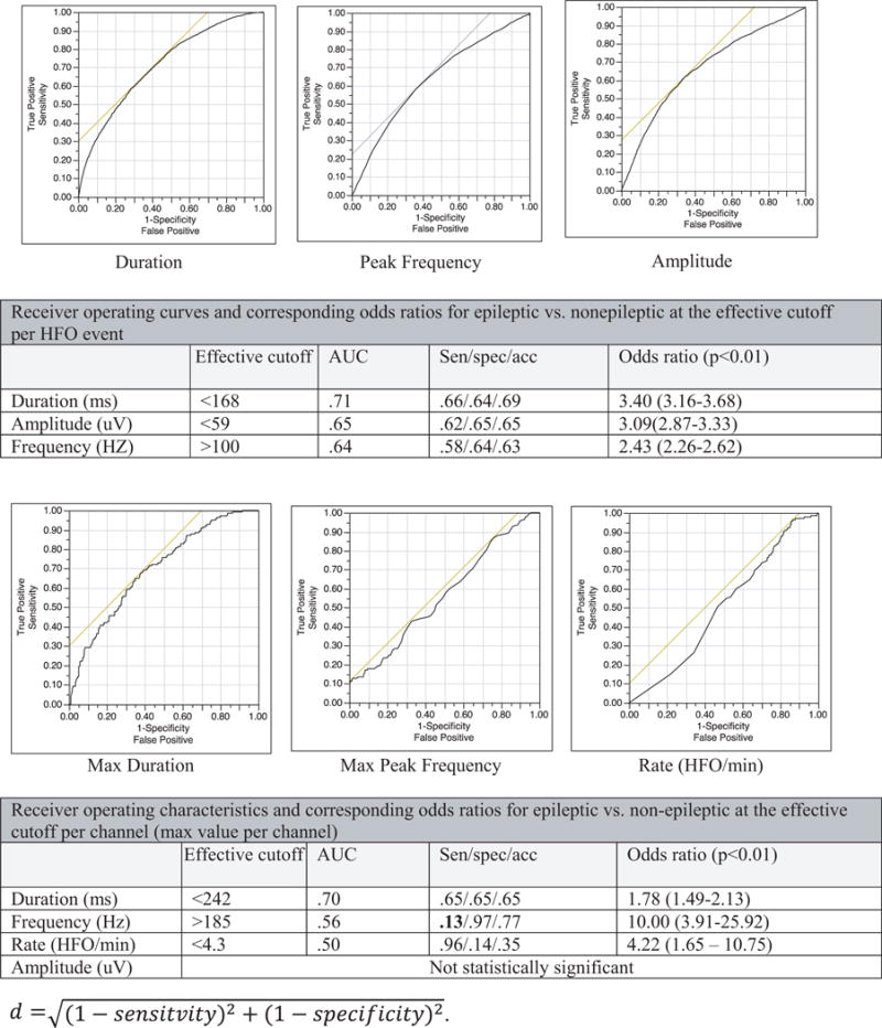
References
-
- Bragin A, Engel J, Jr, Wilson CL, et al. Hippocampal and entorhinal cortex high-frequency oscillations (100–500 Hz) in human epileptic brain and in kainic acid–treated rats with chronic seizures. Epilepsia. 1999;40:127–137. - PubMed
-
- Tatum WO, Dworetzky BA, Freeman WD, et al. Artifact: recording EEG in special care units. J Clin Neurophysiol. 2011;28:264–277. - PubMed
-
- Engel J, Jr, Bragin A, Staba R, et al. High-frequency oscillations: what is normal and what is not? Epilepsia. 2009;50:598–604. - PubMed
Publication types
MeSH terms
Grants and funding
LinkOut - more resources
Full Text Sources
Other Literature Sources
Medical
