. 2014 Dec 2;20(6):1030-7.
doi: 10.1016/j.cmet.2014.11.006.

A parabrachial-hypothalamic cholecystokinin neurocircuit controls counterregulatory responses to hypoglycemia

Affiliations

A parabrachial-hypothalamic cholecystokinin neurocircuit controls counterregulatory responses to hypoglycemia

Alastair S Garfield et al. Cell Metab. .

Abstract

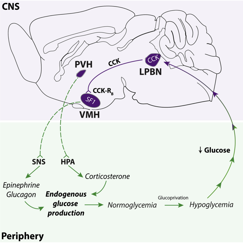
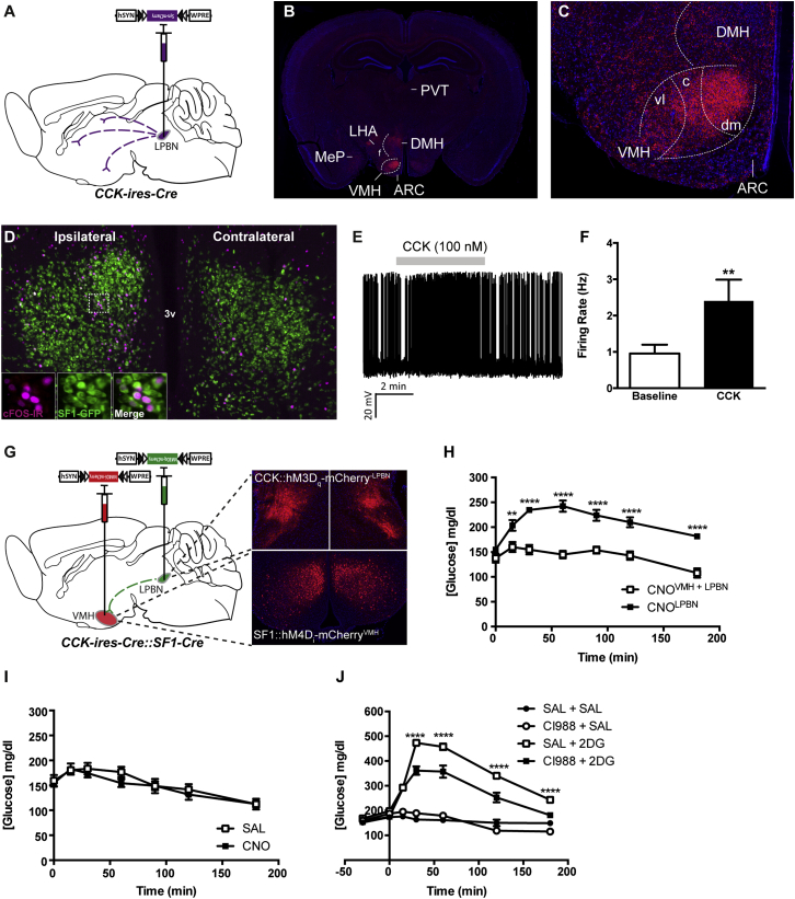
Hypoglycemia engenders an autonomically mediated counterregulatory (CR)-response that stimulates endogenous glucose production to maintain concentrations within an appropriate physiological range. Although the involvement of the brain in preserving normoglycemia has been established, the neurocircuitry underlying centrally mediated CR-responses remains unclear. Here we demonstrate that lateral parabrachial nucleus cholecystokinin (CCK(LPBN)) neurons are a population of glucose-sensing cells (glucose inhibited) with counterregulatory capacity. Furthermore, we reveal that steroidogenic-factor 1 (SF1)-expressing neurons of the ventromedial nucleus of the hypothalamus (SF1(VMH)) are the specific target of CCK(LPBN) glucoregulatory neurons. This discrete CCK(LPBN)→SF1(VMH) neurocircuit is both necessary and sufficient for the induction of CR-responses. Together, these data identify CCK(LPBN) neurons, and specifically CCK neuropeptide, as glucoregulatory and provide significant insight into the homeostatic mechanisms controlling CR-responses to hypoglycemia.


Figures

None
Graphical abstract
Figure 1
Figure 1
CCKLPBN Neurons Are Activated by Glucoprivation (A–D) 2DG- and INS-induced glucoprivation promoted cFOS-IR (red) within the LPBN, as compared to saline controls. Both glucoprivic stimuli elicited cFOS-IR within the sLPBN (B,C), with 2DG also increasing neural activity within the external compartment of the LPBN (B). (D) Quantification of cFOS-IR across the rostral-to-caudal extent of the LPBN (see Figure S1) revealed a significant elevation of cFOS-IR in 2DG- and INS-treated mice above saline controls (n = 3–5 per group; one-way ANOVA, F(2,8) = 31.1, p = 0.0002 with Tukey’s post hoc comparison). (E–G) The sLPBN is located at the rostral and dorsal extreme of the LPBN and defined by the expression of CCK. (F and G) Transgenic labeling of CCK neurons (green) in a CCK-ires-Cre::R26-loxSTOPlox-L10-GFP mouse line recapitulated the known endogenous expression profile. (H–K) 2DG- and INS-induced glucoprivation increased cFOS-IR (red) within CCKLPBN neurons (green) compared to saline controls (white arrows denote colocalized neurons) (n = 3–5 per group; one-way ANOVA, F(2,9) = 23.2, p = 0.0003 with Tukey’s post hoc comparison). (L–N) A subset of CCKLPBN neurons were inhibited by glucose. (L and M) 5/14 CCKLPBN neurons exhibited reversible membrane depolarization in response to a downward glucose step form 5 mM to 0.5 mM (n = 5, repeated-measures one-way ANOVA, F(2,4) = 77.3, p = 0.0003 with Tukey’s post hoc comparison). (N) Representative electrophysiological trace from a spontaneously active glucose-inhibited CCKLPBN neuron. 2DG, 2-deoxyglucose; e, external LPBN; INS, insulin; SAL, saline; scp, superior cerebellar peduncle; s, superior LPBN. All data are presented as mean ± SEM. ∗∗p < 0.01; ∗∗∗p < 0.001.
Figure 2
Figure 2
CCKLPBN Neurons Promote SNS-Mediated CR-Like Responses (A and B) Bilateral stereotaxic injection of Cre-dependent excitatory hM3Dq-mCherry virus into the LPBN of male CCK-ires-Cre mice facilitated real-time activation of CCKLPBN neurons. (A) Representative image of Cre-dependent expression of hM3Dq-mCherry specifically within the LPBN of a CCK-ires-Cre mouse. (B) Membrane potential and firing rate of CCK-ires-Cre::hM3Dq-mCherryLPBN neurons increased upon 5 μM CNO application. (C) CCK-ires-Cre::hM3Dq-mCherryLPBN mice exhibited a significant hyperglycemic response to CNO, compared to saline, administration (n = 7; repeated-measures ANOVA, main effect of treatment [F(1,36) = 39.6, p < 0.0001], main effect of time [F(5,36) = 6.6, p = 0.0002], and interaction [F(5,36) = 4.3, p = 0.003]; post hoc comparisons determined by Sidak’s post hoc test for individual time point analysis). (D–F) CNO treatment evoked an increase in serum glucagon (D; n = 3 per group; t test, t(4) = 26.0, p < 0.0001), corticosterone (E; n = 3 to 4 per group; t test, t(6) = 4.4, p = 0.004), and epinephrine concentrations (F; n = 6–8 per group; t test, t(12) = 2.3, p = 0.04). (G) CCKLPBN neuron activation was associated with increased hepatic G6pc mRNA expression (n = 3 to 4 per group; t test, t(4) = 2.7, p = 0.05) compared to saline controls. (H) CNO-induced hyperglycemia was abolished by pretreatment with pan-specific CCK-receptor antagonist (20 mg/kg proglumide: PROG) (data shown at 60 min after CNO/SAL administration; n = 5; repeated-measures ANOVA, F(4,12) = 5.0, p = 0.04 with Tukey’s post hoc comparison). (I and J) CNO treatment did not influence feeding behavior compared to saline. (I) Three hour dark-cycle food intake (n = 10; paired t test, t(9) = 0.4, p = 0.7) and (J) 3 hr light-cycle food intake (n = 5; paired t test, t(4) = 1.8, p = 0.1) in ad-libitum-fed mice. CNO, clozapine-N-oxide; G6pc, glucose-6-phosphatase; scp, superior cerebellar peduncle; SAL, saline. All data are presented as mean ± SEM; p < 0.05, ∗∗p < 0.01, ∗∗∗p < 0.001, ∗∗∗∗p < 0.0001.
Figure 3
Figure 3
CCKLPBN Neurons Are Necessary for CR-Response to Glucoprivation (A and B) Bilateral stereotaxic injection of Cre-dependent inhibitory hM4Di-mCherry virus into the LPBN of male CCK-ires-Cre mice facilitated the real-time inhibition of CCKLPBN neurons. (A) Representative image of Cre-dependent expression of hM4Di-mCherry specifically within the LPBN of a CCK-ires-Cre mouse. (B) Membrane potential and firing rate of CCK-ires-Cre::hM4Di-mCherryLPBN neurons decreased upon 5 μM CNO application. (C) CNO-induced CCKLPBN neuron silencing in normoglycemic CCK-ires-Cre::hM4Di-mCherryLPBN mice had no effect on blood glucose levels, compared to saline controls (n = 11; repeated-measures ANOVA, main effects of treatment and time, and interaction, not significant). (D and E) CNO-induced CCKLPBN neuron silencing under glucoprivic conditions attenuated the CR-response. (D) 2DG-induced CR was significantly diminished by CNO pretreatment compared to saline (n = 8; repeated-measures ANOVA, main effect of treatment [F(1,49) = 14.3, p < 0.0004], main effect of time [F(6,49) = 64.7, p < 0.0001], and interaction [F(6,49) = 2.8, p = 0.02]; post hoc comparisons determined by Sidak’s post hoc test for individual time point analysis). (E) Likewise, INS-induced CR was significantly diminished by CNO pretreatment compared to saline (n = 5; repeated-measures ANOVA, main effect of treatment [F(1,32) = 4.6, p = 0.04], main effect of time [F(7,32) = 22.3, p < 0.0001], and interaction [F(7,32) = 2.8, p = 0.02]; post hoc comparisons determined by Sidak’s post hoc test for individual time point analysis). Abbreviations: 2DG, 2-deoxyglucose; INS, insulin; scp, superior cerebellar peduncle; SAL, saline. All data are presented as mean ± SEM.; ∗∗p < 0.01; ∗∗∗p < 0.001; ∗∗∗∗p < 0.0001.
Figure 4
Figure 4
CCKLPBN Neurons Engage SF1VMH Neurons to Mediate CR-Responses (A) Unilateral stereotaxic injection of Cre-dependent synpatophysin-mCherry virus into the LPBN of CCK-ires-Cre mice facilitated genetically defined tract tracing of CCKLPBN neuron projections. (B and C) CCKLPBN neurons send ascending projections to the ipsilateral hypothalamus, including the lateral hypothalamus (LH), dorsomedial nucleus (DMH), and VMH. (D) CNO-mediated activation of unilateral CCK-ires-Cre::hM3Dq-mCherryLPBN neurons evokes cFOS-IR (magenta) within ipsilateral SF1VMH neurons (green) in CCK-ires-Cre::SF1-Cre::R26-loxSTOPlox-L10-GFP mice. (E and F) A total of 55% (5/9) of synaptically isolated SF1VMH neurons were activated by CCK (CCK-8S, 100 nM) in ex vivo slice preparations maintained under hypoglycemic conditions (0.5 mM glucose). (E) Representative electrophysiological trace of a SF1VMH neuron demonstrating CCK-induced activation. (F) CCK-responsive SF1VMH neurons exhibited a 2.5-fold increase in firing frequency over baseline upon CCK-8S administration (n = 6, paired t test, t(4) = 4.1, p = 0.01). (G and H) Functional occlusion of CCKLPBN neuron glucoregulation through concomitant silencing of downstream SF1VMH neurons. (H) SF1-Cre::hM4Di-mCherryVMH silencing prevents the CCK-ires-Cre::hM3Dq-mCherryLPBN-mediated CR-response in CNO-treated double transduced mice, as compared to CCK-ires-Cre::hM3Dq-mCherryLPBN only transduced mice (n = 5 per group; two way ANOVA, main effect of treatment [F(1,56) = 188.2, p < 0.0001], main effect of time [F(6,56) = 11.9, p < 0.0001], and interaction [F(6,56) = 4.5, p = 0.0009]; Sidak’s post hoc test for individual time point analysis). (I) SF1VMH neuron silencing does not influence blood glucose concentrations compared to saline (n = 4; repeated-measures ANOVA; main effects of treatment, time, and interaction not significant). (J) The CR-response to 2DG was significantly attenuated by pretreatment with the selective CCKB-receptor antagonist CI988 (n = 5 per group; two-way ANOVA; main effect of treatment [F(3,112) = 369.1, p < 0.0001], main effect of time [F(6,112) = 121.7, p < 0.0001], and interaction [F(18,112) = 36.2, p < 0.001]; post hoc comparisons determined by Tukey’s post hoc test for individual time point analysis). Abbreviations: ARC, arcuate nucleus of the hypothalamus; DMH, dorsomedial nucleus of the hypothalamus; LHA, lateral hypothalamic area; MeP, medial amygdaloid nucleus posterior part; PVT, paraventricular nucleus of the thalamus; SAL, saline; VMH, ventromedial nucleus of the hypothalamus; c, central; vl, ventrolateral; dm, dorsomedial. All data are presented as mean ± SEM; p < 0.05; ∗∗∗p < 0.001; ∗∗∗∗p < 0.0001.

Comment in

References

    1. Alexander G.M., Rogan S.C., Abbas A.I., Armbruster B.N., Pei Y., Allen J.A., Nonneman R.J., Hartmann J., Moy S.S., Nicolelis M.A. Remote control of neuronal activity in transgenic mice expressing evolved G protein-coupled receptors. Neuron. 2009;63:27–39. - PMC - PubMed
    1. Borg W.P., During M.J., Sherwin R.S., Borg M.A., Brines M.L., Shulman G.I. Ventromedial hypothalamic lesions in rats suppress counterregulatory responses to hypoglycemia. J. Clin. Invest. 1994;93:1677–1682. - PMC - PubMed
    1. Borg W.P., Sherwin R.S., During M.J., Borg M.A., Shulman G.I. Local ventromedial hypothalamus glucopenia triggers counterregulatory hormone release. Diabetes. 1995;44:180–184. - PubMed
    1. Briski K.P. Induction of Fos immunoreactivity by acute glucose deprivation in the rat caudal brainstem: relation to NADPH diaphorase localization. Histochem. Cell Biol. 1999;111:229–233. - PubMed
    1. Carter M.E., Soden M.E., Zweifel L.S., Palmiter R.D. Genetic identification of a neural circuit that suppresses appetite. Nature. 2013;503:111–114. - PMC - PubMed

Publication types