. 2015 Feb 16;21(8):3156-66.
doi: 10.1002/chem.201405253. Epub 2014 Dec 3.

NMR hyperpolarization techniques for biomedicine

Affiliations

NMR hyperpolarization techniques for biomedicine

Panayiotis Nikolaou et al. Chemistry. .

Abstract

Recent developments in NMR hyperpolarization have enabled a wide array of new in vivo molecular imaging modalities, ranging from functional imaging of the lungs to metabolic imaging of cancer. This Concept article explores selected advances in methods for the preparation and use of hyperpolarized contrast agents, many of which are already at or near the phase of their clinical validation in patients.

Keywords: dynamic nuclear polarization; hyperpolarization; medicinal chemistry; molecular imaging; spin exchange optical pumping.


Figures

Figure 1
Figure 1
The process of Dynamic Nuclear Polarization (DNP) using high-power microwave irradiation of electron spins at low temperatures (few Kelvin) and high magnetic field (several Tesla). (a) Glass matrix containing 13C-labeled metabolite and radicals with unpaired electron is formed inside the high-field DNP magnet at low temperature. (b) High-power microwave irradiation at the electron resonance frequency enables 13C hyperpolarization via polarization transfer from free electrons. (iii) A long-lived 13C hyperpolarized state is prepared.
Figure 2
Figure 2
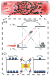
Spin-Exchange Optical Pumping.[9] (a) SEOP requires an optical cell containing a noble gas, buffer gases (e.g. N2, as shown), and a small quantity of vaporized alkali metal (typically Rb or Cs[47]), irradiated by laser light resonant with an absorption line of the alkali metal (e.g. 794.8 nm for Rb D1 transition). Collisions with N2 non-radiatively quench excited Rb states, effectively suppressing deleterious Rb fluorescence that can depolarize other Rb spins.[48] The first step of SEOP involves the absorption of photons of the same circular polarization, which conserves angular momentum by selectively depleting population from one of two Rb ground electronic (mJ=±1/2) states (neglecting Rb nuclear spin for simplicity). Collisions with other gas-phase species tend to equalize the excited-state populations and the ground states are repopulated at effectively equal rates. However, since only one ground state is depleted by the laser, ground-state population accumulates on the other mJ state, leaving the Rb electronically spin-polarized; a weak magnetic field along the direction of laser propagation (not shown) helps to maintain the electron spin polarization. Gas-phase collisions also allow spin exchange (c) between the polarized Rb electron spins and the noble gas nuclear spins, a process mediated by Fermi-contact hyperfine interactions. Constant laser illumination of the Rb vapor therefore allows the nuclear spin polarization to accumulate over time, thereby generating the hyperpolarized noble gas.
Figure 3
Figure 3
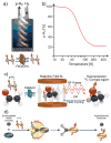
a–b) Conversion of ‘normal’ H2 gas (75% ortho- and 25% para-isomers) into parahydrogen (p-H2). a) Schematic of a p-H2 generator, where the entering room-temperature (‘normal’) H2 gas is cryo-cooled to ≤77 K and catalytically converted under local equilibrium to a mixture of spin isomers that preferentially favors p-H2. Because the catalyst is confined to a cryogenically cooled chamber of the polarizer, once the p-H2 leaves the chamber it is kinetically trapped in the para-state. b) Temperature dependence of the equilibrium p-H2 percentage. Liquid N2 temperature (77 K) allows for preparation of 50% p-H2, whereas temperatures below 20 K enable production of >97% p-H2 fraction. c) The process of Parahydrogen Induced Polarization (PHIP). The pairwise addition of a p-H2 molecule to a molecular precursor, which is typically accomplished across a C=C or C≡C bond adjacent to a 13C nucleus using a hetero- or homogeneous catalyst. The resulting chemically ‘unlocked’ nuclear spin hyperpolarization of the nascent, magnetically inequivalent protons can be used as-is, or transferred to the typically longer-lived (greater T1) 13C site using either a RF pulse sequence or a field-cycling method. d) The process of Signal Amplification by Reversible Exchange (SABRE). A metal complex [M] enables p-H2 and a substrate to be transiently co-located under conditions of dynamic exchange, resulting in spontaneous polarization transfer[11, 62] from p-H2 to the substrate.
Figure 4
Figure 4
MR detection concepts of HP contrast agents. a) In vivo administration of HP 1-13C-pyruvate leads to its in vivo uptake as well as its subsequent metabolism to 1-13C-alanine (using alanine transaminase [ALT]), 1-13C-lactate (using lactate dehydrogenase [LDH]), which can be differentiated using 13C chemical shifts, and b) detected using Chemical Shift Imaging (CSI) to simultaneously produce multiple metabolic maps. Note the red (lactate), blue (alanine) and green (pyruvate) color coding in a) and b). c) Trigonometric dependence of observed (sine) and remaining (cosine) magnetization as a function of RF pulse excitation (or “tipping”) angle. d) Fundamental blocks of RF pulse sequences for conventional MR (top); hyperpolarized (HP) MR with fixed small-angle encoding pulses (middle), and HP MR with variable-angle encoding pulses (bottom).
Figure 5
Figure 5
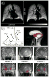
Examples of biomedical use of hyperpolarized contrast agents in human subjects for pulmonary functional imaging,[5b] spectroscopy and imaging of brain uptake of 129Xe,[79] and molecular imaging of prostate cancer.[27] a) Two slices selected from a 3D 129Xe GRE chest image from a healthy human volunteer following inhalation of HP Xe from an 800-cc Tedlar bag containing a mixture of HP Xe (86%-enriched 129Xe) : 35% N2, 65 : 35. Subject performed two respiration cycles (total lung capacity to functional residual capacity), inhaled from bag, and then took a small gulp of air (to help push HP Xe out of the trachea); TE/TR, 1.12/11 ms (specific absorption rate-limited); tipping angle, 6°; 80 × 80 × 14; acquisition time, 4.5 s; FOV, 320 × 320 × 196 mm3; 2 × 2 × 14 mm3 digital resolution after zero-filling (SNR ~8–15)[5b] b) Left: HP 129Xe spectrum from the brain of a healthy human subject following inhalation of HP Xe from a 1 L Tedlar bag containing Xe : N2 gas mixture (85 : 15 by volume) (129Xe enrichment: 87%; PXe~60%). Tentative assignment: 214.5 ppm: red blood cells (RBCs); 198 ppm: blood plasma; 195.3 ppm: grey matter; 192.5 ppm: white matter; 187 ppm: lipid (15 scans; TR=2 s; pulse tipping angle: ~55–65º). Right: sagittal 129Xe MR image (false color) of a healthy human subject overlayed on a corresponding greyscale 1H image following inhalation of HP Xe (same as above, but 100% Xe in bag). 129Xe image shows the signal from the Xe/RBC resonance alone (214.5±0.5 ppm; 2D pulse-acquire; 8X8 matrix of FIDs (zero-filled to 32×32) with 256 points; FOV = 30 cm; slice thickness = 20 cm; pulse tipping angle: ~20º; TR=0.3 s; total 129Xe imaging time: ~20 s). Figures generously provided by Madhwesha Rao, U. Sheffield, UK.[79] c) Representative examples of 3D single-time-point Magnetic Resonance Spectroscopic Imaging (MRSI) data of three prostate cancer subjects after IV injection of HP 1-13C-pyruvate.[27] The axial T2-weighted images and false-color overlays of hyperpolarized 1-13C-lactate/1-13C-pyruvate ratio are from the three patients labeled as B, C, and D. All three of the patients had biopsy-proven Gleason grade 3 + 3 prostate cancer and received the highest dose of hyperpolarized 1-13C-pyruvate (0.43 ml/kg). Patients B, C, and D had current PSAs of 5.1, 9.8, and 1.9 ng/ml, respectively. The SNR and metabolite ratios in the regions highlighted in color on the image overlays are given in Table 2 of Ref. [27] From S. J. Nelson, et al., Sci. Transl. Med. 2013, 5, 198ra108. Reprinted with permission from AAAS.

Similar articles

Cited by

References

    1. Zeeman P. Nature. 1897;55:347.
    1. Carver TR, Slichter CP. Phys Rev. 1953;92:212–213.
    1. Nikolaou P, Coffey AM, Barlow MJ, Rosen M, Goodson BM, Chekmenev EY. Anal Chem. 2014;86:8206–8212. - PMC - PubMed
    1. Kurhanewicz J, Vigneron DB, Brindle K, Chekmenev EY, Comment A, Cunningham CH, DeBerardinis RJ, Green GG, Leach MO, Rajan SS, Rizi RR, Ross BD, Warren WS, Malloy CR. Neoplasia. 2011;13:81–97. - PMC - PubMed
    1. Muradyan I, Butler JP, Dabaghyan M, Hrovat M, Dregely I, Ruset I, Topulos GP, Frederick E, Hatabu H, Hersman WF, Patz S. J Magn Reson Imaging. 2013;37:457–470. - PMC - PubMed
    2. Nikolaou P, Coffey AM, Walkup LL, Gust BM, Whiting N, Newton H, Barcus S, Muradyan I, Dabaghyan M, Moroz GD, Rosen M, Patz S, Barlow MJ, Chekmenev EY, Goodson BM. Proc Natl Acad Sci U S A. 2013;110:14150–14155. - PMC - PubMed

Publication types

MeSH terms