. 2014 Dec 3;34(49):16309-19.
doi: 10.1523/JNEUROSCI.4622-13.2014.

Actions of NPY, and its Y1 and Y2 receptors on pulsatile growth hormone secretion during the fed and fasted state

Affiliations

Actions of NPY, and its Y1 and Y2 receptors on pulsatile growth hormone secretion during the fed and fasted state

Lili Huang et al. J Neurosci. .

Abstract

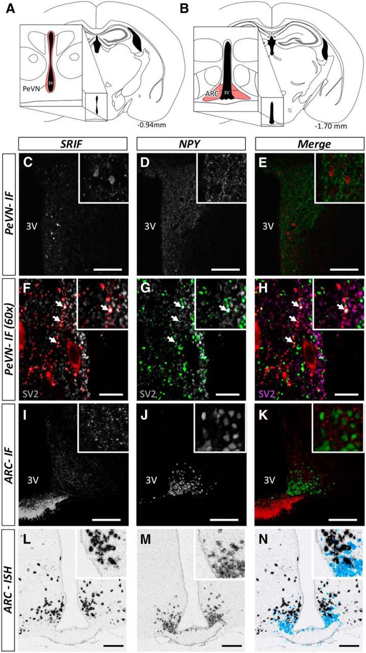
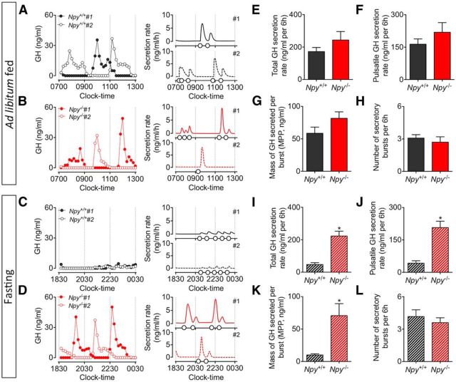
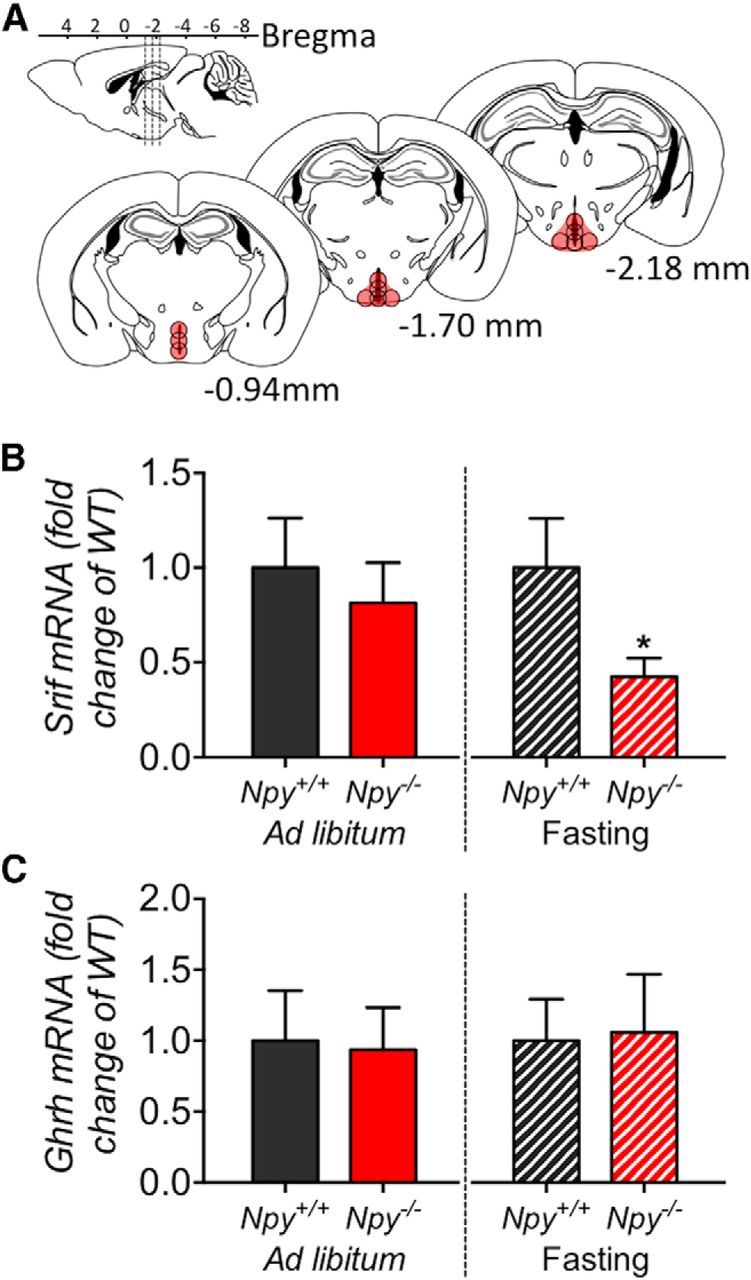
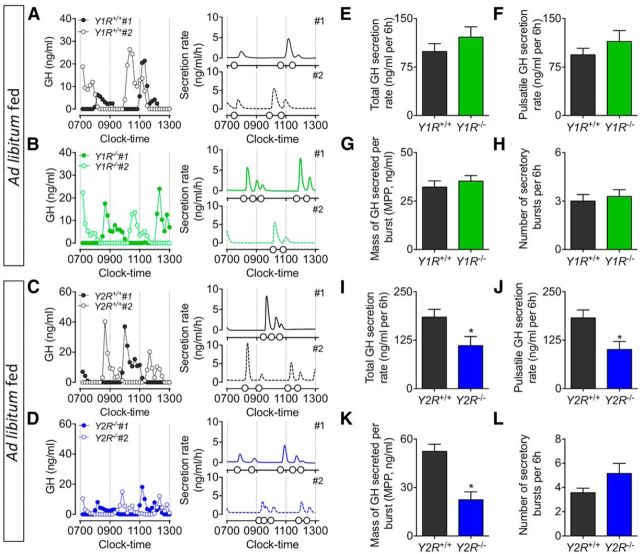
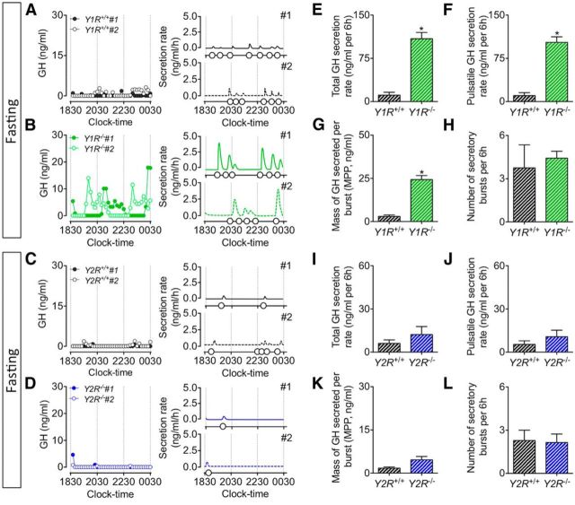
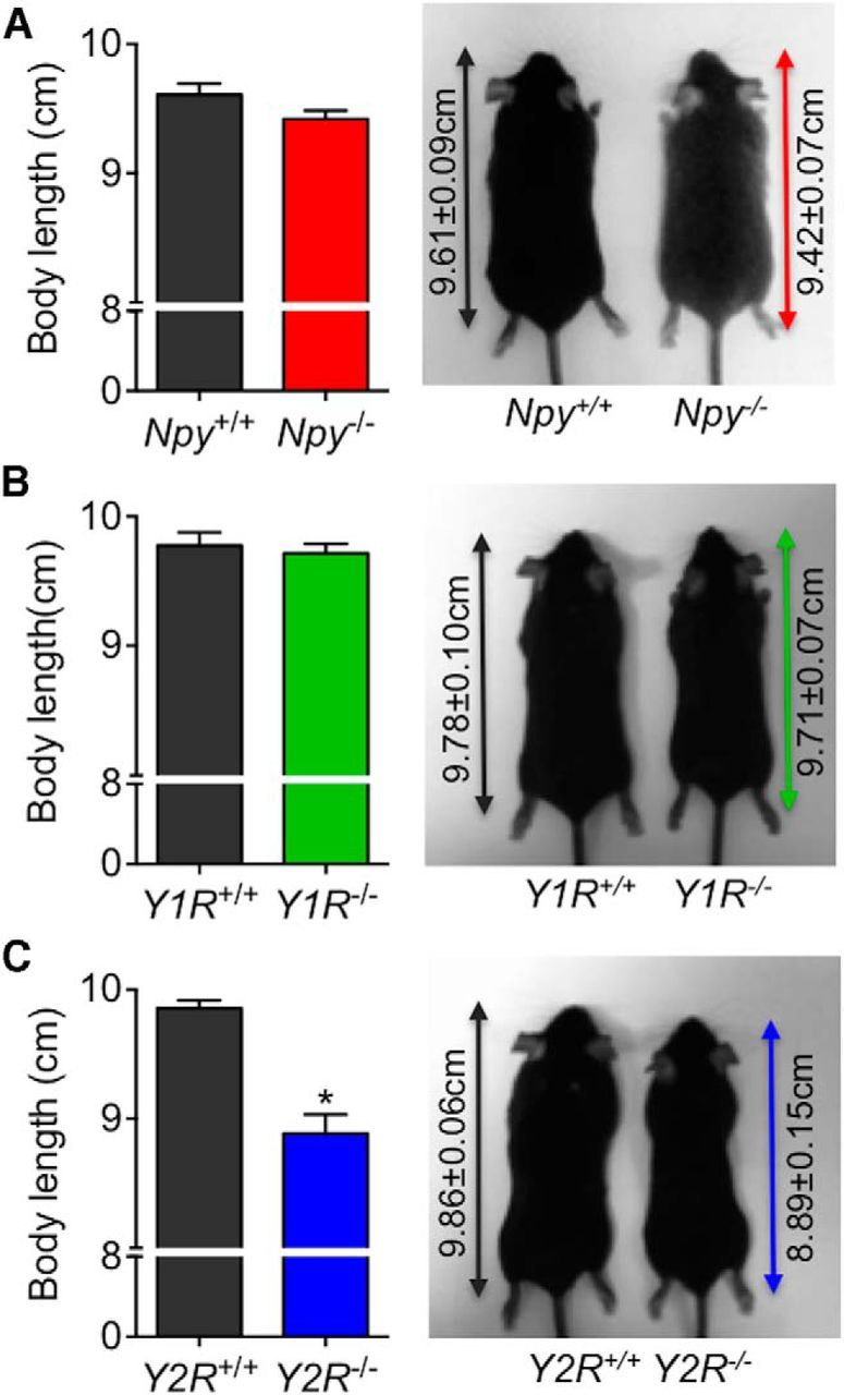
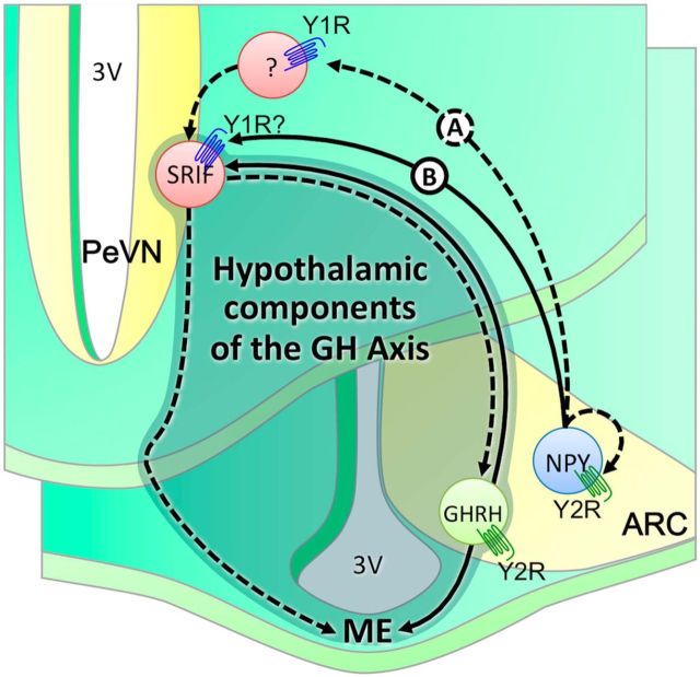
The hypothalamic NPY system plays an important role in regulating food intake and energy expenditure. Different biological actions of NPY are assigned to NPY receptor subtypes. Recent studies demonstrated a close relationship between food intake and growth hormone (GH) secretion; however, the mechanism through which endogenous NPY modulates GH release remains unknown. Moreover, conclusive evidence demonstrating a role for NPY and Y-receptors in regulating the endogenous pulsatile release of GH does not exist. We used genetically modified mice (germline Npy, Y1, and Y2 receptor knock-out mice) to assess pulsatile GH secretion under both fed and fasting conditions. Deletion of NPY did not impact fed GH release; however, it reversed the fasting-induced suppression of pulsatile GH secretion. The recovery of GH secretion was associated with a reduction in hypothalamic somatotropin release inhibiting factor (Srif; somatostatin) mRNA expression. Moreover, observations revealed a differential role for Y1 and Y2 receptors, wherein the postsynaptic Y1 receptor suppresses GH secretion in fasting. In contrast, the presynaptic Y2 receptor maintains normal GH output under long-term ad libitum-fed conditions. These data demonstrate an integrated neural circuit that modulates GH release relative to food intake, and provide essential information to address the differential roles of Y1 and Y2 receptors in regulating the release of GH under fed and fasting states.

Keywords: NPY; NPY-receptors; fasting; feeding; growth hormone; somatostatin.


Figures

Figure 1.
Figure 1.
Illustration of anatomical interactions between SRIF and NPY-expressing neurons within the hypothalamus (A, B) by immunofluorescence (C–K) and in situ hybridization (L–N). Brain slices were collected between −0.94 and −1.70 bregma. Schematic representation of specific areas within the hypothalamus, representative of the PeVN (A, enlarged inset, shaded areas in red) and ARC (B, enlarged inset, shaded area in red). Within the PeVN (C–H) we observed a number of SRIF-immunopositive neurons (red) in close apposition with NPY-GFP-immunopositive fibers (green; C–E). Further assessment using SV2 (gray; F and G; purple; H) revealed the synaptic interactions between SRIF and NPY-positive fibers (F–H; colocalization is indicated by white arrows). SRIF-positive immunoreactivity and NPY-GFP within the ARC (I–K) demonstrate punctuate SRIF expression, distributed among NPY-positive GFP neuronal cell bodies. In situ hybridization illustrates the proportion of Srif (black) and Npy (light blue) mRNA-expressing neurons within the ARC (L–N). These two populations of neurons were in close proximity. Scale bars: C–E, I–N, 100 μm; F–H, 10 μm. Inserts illustrate a magnified view of the figures. Representative images illustrate interactions verified across four animals.
Figure 2.
Figure 2.
Representative examples of pulsatile GH secretion profiles (2 animals; #1, closed circles; #2, open circles; A–D, left) and corresponding output figures (A–D, right) in male adult Npy−/− mice (red) and age-matched wild-type littermates (black). Assessment of pulsatile GH secretion was done in ad libitum-fed mice (A, B) and during the first 6 h of fasting (C, D). Samples were collected for 6 h at 10 min intervals, starting at 0700 h (fed) and 1830 h (fasting). Corresponding output figures illustrate the onset of GH pulses (pulse onset indicated by open circles along the x-axis). GH secretory parameters following deconvolution analysis are illustrated in E–H (ad libitum fed) and I–L (fasting). Total (E) and pulsatile GH secretion rate (F), the mass of GH secreted per burst (G), and number of secretory bursts (H) in Npy−/− mice were comparable to ad libitum-fed wild-type mice. In contrast, the fasting-induced suppression of GH secretion observed in wild-type mice was prevented in Npy−/− mice. This was characterized by a significant increase in total (I) and pulsatile GH secretion rate (J), and the mass of GH secreted per burst (K) in Npy−/− mice. The number of secretory bursts (L) remained unchanged. Data are presented as mean ± SEM, N = 10–12, *p < 0.05.
Figure 3.
Figure 3.
Comparison of Ghrh and Srif mRNA expression within the ARC/PeVN of Npy−/− mice and corresponding WT mice under ad libitum-fed conditions and following 6 h of fasting. Micropunch biopsies of tissue representing the ARC/PeVN (between −0.94 and −2.18 bregma) were collected from frozen brain sections (A, location of punch biopsies illustrated in red). Compared with wild-type mice, deletion of NPY did not result in a significant difference in Srif (B, left) or Ghrh (C, left) mRNA expression under ad libitum-fed conditions. In contrast, deletion of NPY resulted in a reduction of Srif mRNA expression (B, right) following 6 h of fasting. Ghrh mRNA expression remained unchanged (C, right). Data are presented as mean ± SEM, N = 5–6, *p < 0.05.
Figure 4.
Figure 4.
Assessment of circulating levels of blood glucose, NPY, and PYY in response to fasting. Glucose was measured every 15 min for a duration of 150 min, and corrected to starting levels (A). Circulating levels of NPY (B) and PYY (C) were determined following 45 and 90 min of fasting. Fasting resulted in a decline in circulating levels of glucose by 45 min of food withdrawal (A). Unlike glucose, circulating levels of NPY and PYY remained unchanged for the duration of the fasting period (B, C). Data are presented as mean ± SEM, N = 12, *p < 0.05.
Figure 5.
Figure 5.
Representative examples of pulsatile GH secretion profiles (2 animals; #1, closed circles; #2, open circles; A–D, left) and corresponding output figures (A–D, right) from ad libitum-fed male adult mice with germline deletion of Y1 (Y1R−/−; green, B) or Y2 (Y2R−/−; blue, D) receptors and age-matched littermates (black, A and C) under ad libitum-fed conditions. Samples were collected for 6 h at 10 min intervals, starting at 0700 h. Corresponding output figures illustrate the onset of GH pulses (pulse onset indicated by open circles along the x-axis). GH secretory parameters following deconvolution analysis are illustrated in E–H (Y1R−/−) and I–L (Y2R−/−). Total (E) and pulsatile GH secretion rate (F), the mass of GH secreted per burst (G), and number of secretory bursts (H) in Y1R−/− mice were comparable to ad libitum-fed wild-type mice. GH secretion was significantly reduced in Y2R−/− mice as characterized by a significant reduction in total (I), pulsatile (J), and the mass of GH secreted per burst (K). While not significant, the number of secretory bursts (L) was elevated in Y2R−/− mice (p = 0.06). Data are presented as mean ± SEM, N = 4–7, *p < 0.05.
Figure 6.
Figure 6.
Representative examples of pulsatile GH secretion profiles (2 animals; #1, closed circles; #2, open circles; A–D, left) and corresponding output figures (A–D, right) from male adult mice with germline deletion of the Y1 (Y1R−/−; green, B) and Y2 (Y2R−/−; blue, D) receptor and age-matched littermates (black, A and C) following 6 h of food withdrawal. Samples were collected for 6 h at 10 min intervals, starting at 1830 h. Corresponding output figures illustrate the onset of GH pulses (pulse onset indicated by open circles along the x-axis). GH secretory parameters following deconvolution analysis are illustrated in E–H (Y1R−/−) and I–L (Y2R−/−). Deletion of Y1R resulted in a prevention of the fasting-induced suppression of pulsatile GH release, and was characterized by an increase in total (E) and pulsatile GH secretion rate (F), and the mass of GH secreted per burst (G). In contrast, pulsatile GH release was suppressed in fasting Y2R−/− mice. This was characterized by a comparable total (I) and pulsatile GH secretion (J), and the mass of GH secreted per burst (K) when compared with age-matched fasting wild-type littermates. Data are presented as mean ± SEM, N = 4–7, *p < 0.05.
Figure 7.
Figure 7.
Body length and representative images of adult mice following germline deletion of NPY (Npy−/−; A), Y1R (Y1R−/−; B), and Y2R (Y2R−/−; C). Body length in Npy−/− and Y1R−/− mice was comparable to the age-matched wild-type mice, whereas Y2R−/− mice were significantly shorter compared with age-matched wild-type littermates. Body length was measured from nose to rump as indicated by the double arrow line. Data are presented as mean ± SEM, N = 4–12, *p < 0.05.
Figure 8.
Figure 8.
Schematic representation defining interactions between hypothalamic components of the GH axis (shaded), NPY neurons, and Y-receptors. Fasting induces the activation of NPY neurons in the ARC. Current and published observations suggest that altered NPY activity promotes SRIF activity, presumably through activation of the postsynaptic Y1 receptors. Acting through intermediate inhibitory neurons, the Y1R may stimulate SRIF-ergic tone through withdrawal in inhibition (Scenario A). Alternatively, Y1R may directly modulate SRIF-egic tone via nonclassical G-protein-mediated pathways (Scenario B). In both scenarios, increased SRIF-ergic tone will inhibit GHRH-induced GH release, or directly inhibit somatotroph activity and thus GH release. Accordingly, GH release is not suppressed in fasting mice following germline deletion of NPY or Y1R. Under ad libitum-fed conditions, activation and/or inactivation of NPY neurons in the ARC are under tight control of Y2 receptors. Deletion of Y2 receptors results in impaired peak GH release, presumably through a potential increase in NPY feedback to SRIF neurons resulting in an increase in SRIF-ergic tone. Of interest, GHRH neurons express the Y2R (Lin et al., 2007) and promote GHRH activity (Osterstock et al., 2010). Thus, germline loss of Y2R may directly impact GHRH activity in ad libitum-fed mice, and thus GHRH-mediated GH release. PVN, paraventricular nucleus; 3V, third ventricle; ME, medium eminence. Dashed lines represent an inhibitory effect whereas solid lines represent a stimulatory effect.

References

    1. Adams EF, Venetikou MS, Woods CA, Lacoumenta S, Burrin JM. Neuropeptide Y directly inhibits growth hormone secretion by human pituitary somatotropic tumours. Acta Endocrinol. 1987;115:149–154. - PubMed
    1. Attanasio AF, Shalet SM. Growth hormone and the transition from puberty into adulthood. Endocrinol Metab Clin North Am. 2007;36:187–201. doi: 10.1016/j.ecl.2006.11.002. - DOI - PubMed
    1. Balasubramaniam A, Joshi R, Su C, Friend LA, James JH. Neuropeptide Y (NPY) Y2 receptor-selective agonist inhibits food intake and promotes fat metabolism in mice: combined anorectic effects of Y2 and Y4 receptor-selective agonists. Peptides. 2007;28:235–240. doi: 10.1016/j.peptides.2006.08.041. - DOI - PubMed
    1. Baldock PA, Sainsbury A, Couzens M, Enriquez RF, Thomas GP, Gardiner EM, Herzog H. Hypothalamic Y2 receptors regulate bone formation. J Clin Invest. 2002;109:915–921. doi: 10.1172/JCI14588. - DOI - PMC - PubMed
    1. Batterham RL, Cowley MA, Small CJ, Herzog H, Cohen MA, Dakin CL, Wren AM, Brynes AE, Low MJ, Ghatei MA, Cone RD, Bloom SR. Gut hormone PYY3–36 physiologically inhibits food intake. Nature. 2002;418:650–654. doi: 10.1038/nature00887. - DOI - PubMed

Publication types

LinkOut - more resources