. 2014 Nov 29:15:54.
doi: 10.1186/s12865-014-0054-z.

Orphan nuclear receptor Nur77 Inhibits Oxidized LDL-induced differentiation of RAW264.7 murine macrophage cell line into dendritic like cells

Affiliations

Orphan nuclear receptor Nur77 Inhibits Oxidized LDL-induced differentiation of RAW264.7 murine macrophage cell line into dendritic like cells

Liu-Hua Hu et al. BMC Immunol. .

Abstract

Background: Nur77 is an orphan nuclear receptor expressed in human atheroma. In vascular cells in vitro, Nur77 expression is induced by pro-inflammatory factors, such as oxidized LDL (oxLDL).

Methods: We analyze the role of Nur77 in the oxLDL-induced differentiation of macrophages into dendritic cells (DC). The murine RAW264.7 macrophage cell line was stably transfected with expression plasmids encoding either GFP or GFP fusions with either full-length Nur77 (GFP-Nur77), Nur77 lacking the DNA binding domain (GFP-Nur77-ΔDBD) or Nur77 lacking the transactivation domain (GFP-Nur77-ΔTAD).

Results: GFP-Nur77 overexpression significantly suppressed the effect of oxLDL treatment on DC morphologic changes, expression of DC maturation markers, endocytic activity, allogeneic activation of T cell proliferation, and the activity and secretion of pro-inflammatory cytokines. Analysis of GFP-Nur77-ΔTAD and GFP-Nur77-ΔDBD indicated that the Nur77 DNA binding and transactivation domains were both required for this effect. GFP-Nur77-ΔDBD consistently had the opposite effect to GFP-Nur77, increasing DC-type differentiation in all assays. Interestingly, GFP-Nur77-ΔDBD protein was cytosolic, whereas GFP-Nur77 and GFP-Nur77-ΔTAD were both nuclear.

Conclusions: These data show that GFP-Nur77 inhibited differentiation of oxLDL-treated macrophages into DC. The effects of Nur77 on the macrophage phenotype may involve changes in its subcellular distribution.


Figures

Figure 1
Figure 1
Characterization of stable RAW264.7 cell lines expressing Nur77 and Nur77 deletion mutants. (A) Schematic structure of the Nur77 gene and deletion mutants lacking either the transactivation domain (TAD) or DNA binding domain (DBD). (B) Expression of Nur77 protein in untransfected RAW264.7 cells and in RAW264.7 cell lines expressing GFP or GFP-Nur77. Expression of GFP-Nur77 is 3–4-fold higher than endogenous Nur77 in untransfected or GFP-transfected cells. (C) Expression of GFP-Nur77-ΔTAD and GFP-Nur77-ΔDBD fusion proteins in stably transfected RAW264.7 cells. β-actin expression was used to control for protein loading. A representative of three separate experiments is shown. (D) Subcellular localization of GFP-Nur77, GFP-Nur77-ΔTAD, and GFP-Nur77-ΔDBD in RAW264.7 cells.
Figure 2
Figure 2
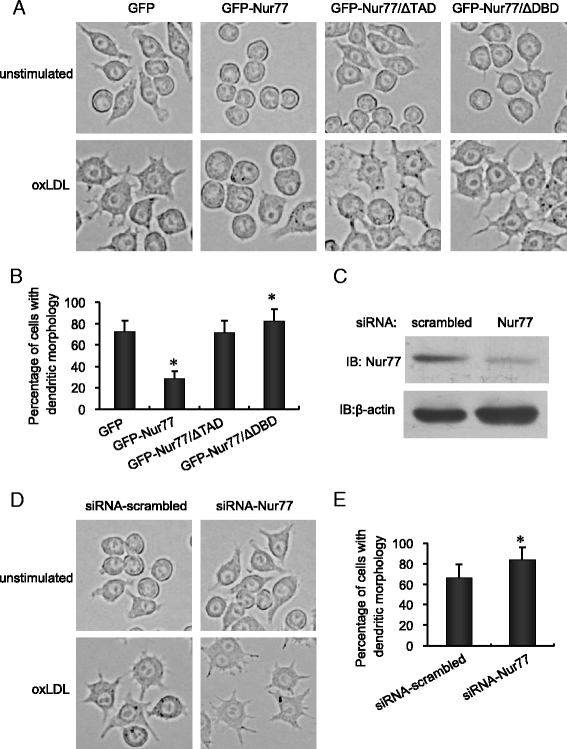
Nur77 inhibits DC morphological changes in oxLDL-treated RAW264.7 cells. (A) RAW264.7 cells stably expressing GFP-Nur77, GFP-Nur77-ΔTAD, or GFP-Nur77-ΔDBD were treated with oxLDL (10 μg/ml) for 48 h and visualized by phase contrast microscopy (400×). Results are representative of three separate experiments. (B) Cells with DC morphology were calculated as the percentage of all cells observed in 10 different fields at 400× magnification. The bars represent mean ± SD from three experiments. *p <0.05 compared with GFP-expressing control. (C) Western blots showing endogenous Nur77 in RAW264.7 cells 48 h after transfection with either scrambled siRNA or Nur77 siRNA. Similar results were obtained in three separate experiments. (D) Phase contrast images showing the morphology of oxLDL-treated RAW264.7 cells transfected with either scramble siRNA or Nur77 siRNA. Cultured cells were visualized by phase contrast microscopy as described in (A) and cells with DC morphology were calculated as described in (B). (E) *p <0.05 compared with scrambled control.
Figure 3
Figure 3
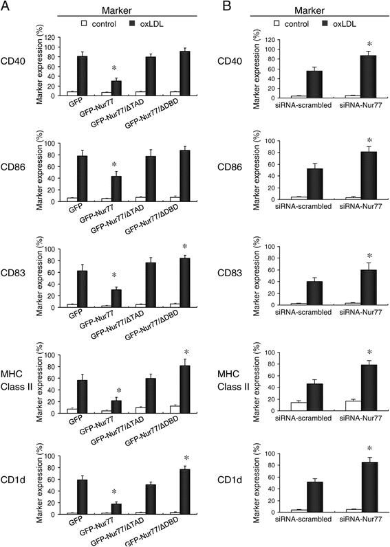
Flow cytometry analysis of the cell surface phenotype of oxLDL-treated RAW264.7 cell lines. (A) Expression of CD40, CD86, CD83, MHC Class II, and CD1d in RAW264.7 cells lines treated with oxLDL for 48 h. Mean ± SD of three independent experiments is shown. *p <0.05 compared with GFP-expressing control. (B) Expression of CD40, CD86, CD83, MHC Class II, and CD1d in RAW264.7 cells transfected with either scrambled siRNA or Nur77 siRNA and stimulated with oxLDL for 48 h. *p <0.05 compared with scrambled control.
Figure 4
Figure 4
Nur77 enhanced the endocytic activity but suppressed the DNA synthesis of RAW264.7 treated by oxLDL. (A) Flow cytometry analysis of endocytic activity using lucifer yellow (LY) uptake by oxLDL-treated RAW264.7 cells stably expressing GFP-Nur77, GFP-Nur77-ΔTAD, or GFP-Nur77-ΔDBD. Mean ± SD of three independent experiments is shown. *p <0.05 compared with GFP-expressing control. (B) Flow cytometry analysis of BrdU incorporation by co-cultures of allogeneic T cells and RAW264.7 cells stably expressing either GFP, GFP-Nur77, GFP-Nur77-ΔTAD, or GFP-Nur77-ΔDBD. Mean ± SD of three independent experiments is shown. *p <0.05 compared with GFP-expressing control.
Figure 5
Figure 5
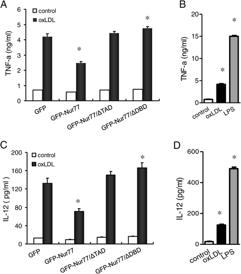
Nur77 overexpression in RAW264.7 cells reduces oxLDL-induced inflammatory cytokine synthesis. Sandwich ELISA analysis of TNF-α (A) and IL-12 (C) for the supernatants of oxLDL-treated RAW264.7 cells stably expressing GFP, GFP-Nur77, GFP-Nur77-ΔTAD or GFP-Nur77-ΔDBD. Data represent mean ± SD of three independent experiments for 1 × 106 cells, *p <0.05 compared with GFP-expressing control cell. (B) and (D) showed the level of TNF-α and IL-12 in GFP-expressing control cells with or without stimulated by oxLDL or LPS, respectively. The bars represent mean ± SD from three experiments. *p <0.05 compared with control cells.

References

    1. Dahl TB, Arne Y, Mona S, Erik O, Arve D, Annika M, Damås JK, Tunheim SH, Thor U, Camilla S, Bjørn B, Serena T, Lars G, Frøland SS, Kirsten K-S, David R, Pål A, Bente H. Increased Expression of Visfatin in Macrophages of Human Unstable Carotid and Coronary Atherosclerosis: Possible Role in Inflammation and Plaque Destabilization. Circulation. 2007;115(8):972–980. doi: 10.1161/CIRCULATIONAHA.106.665893. - DOI - PubMed
    1. Parks BW, Lusis AJ. Macrophage accumulation in atherosclerosis. N Engl J Med. 2013;369(24):2352–2353. doi: 10.1056/NEJMcibr1312709. - DOI - PMC - PubMed
    1. Frostegård J. Immunity, atherosclerosis and cardiovascular disease. BMC Med. 2013;11:117. doi: 10.1186/1741-7015-11-117. - DOI - PMC - PubMed
    1. Yuri V. Bobryshev. Dendritic cells and their role in atherogenesis. Lab Invest. 2010;90:970–984. doi: 10.1038/labinvest.2010.94. - DOI - PubMed
    1. Shen LH, Zhou L, Wang BY, Pu J, Hu LH, Chai DJ, Wang L, Zeng JZ, He B. Oxidized low-density lipoprotein induces differentiation of RAW264.7 murine macrophage cell line into dendritic-like cell. Atherosclerosis. 2008;199:257–264. doi: 10.1016/j.atherosclerosis.2007.12.002. - DOI - PubMed

Publication types

MeSH terms

Substances

LinkOut - more resources