

Contour changes in human alveolar bone following tooth extraction of the maxillary central incisor
- PMID: 25471836
- PMCID: PMC4265561
- DOI: 10.1631/jzus.B1400119
Contour changes in human alveolar bone following tooth extraction of the maxillary central incisor
Abstract
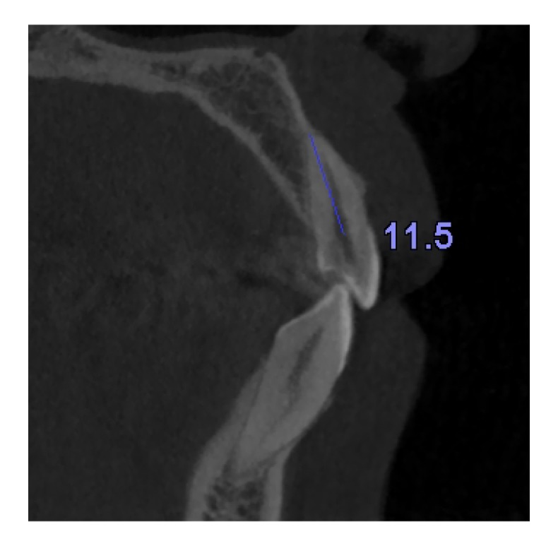
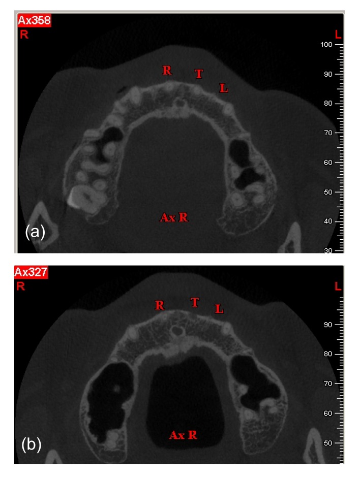
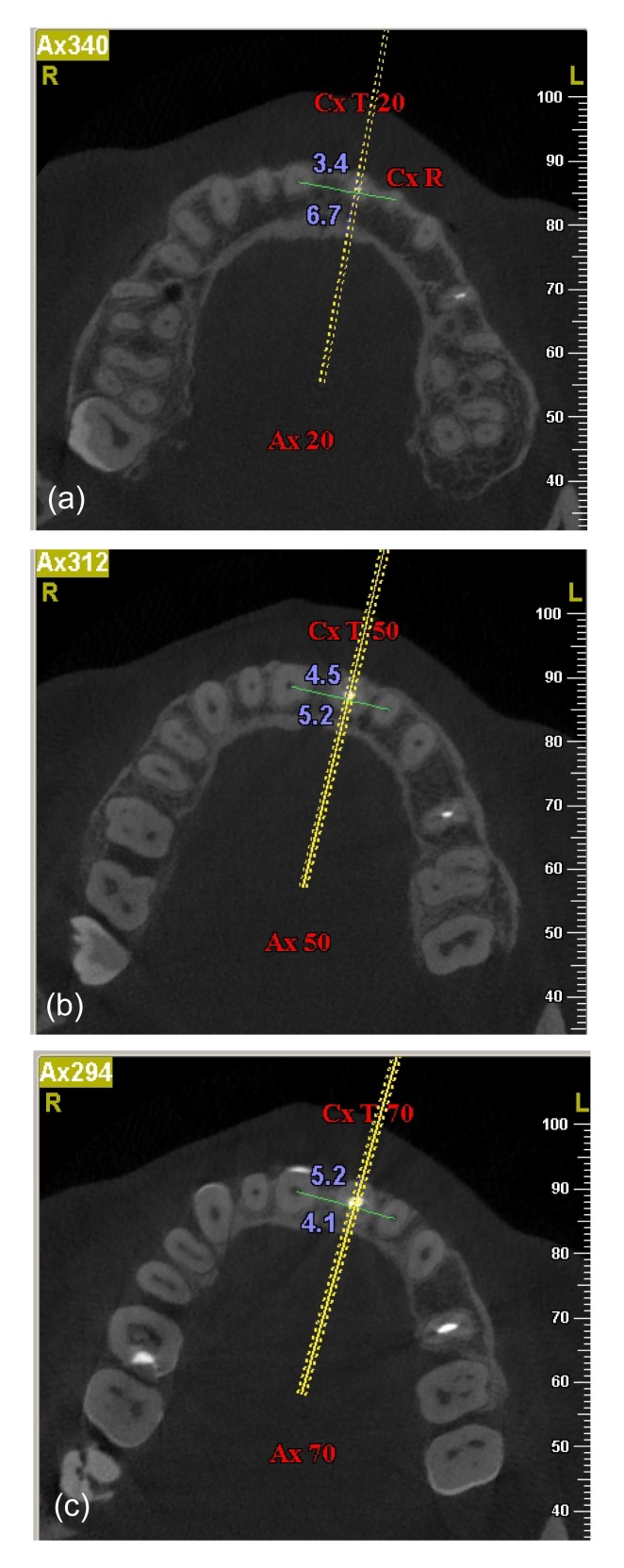
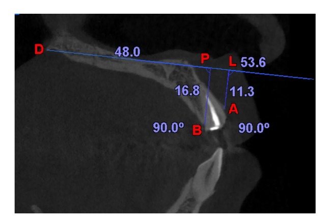
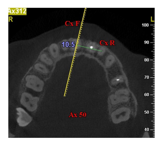
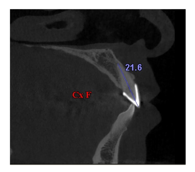
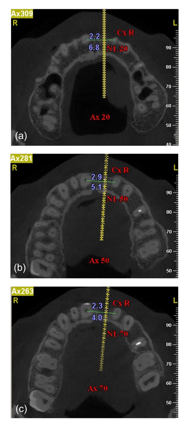
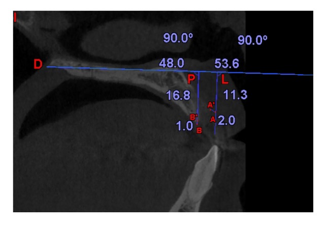
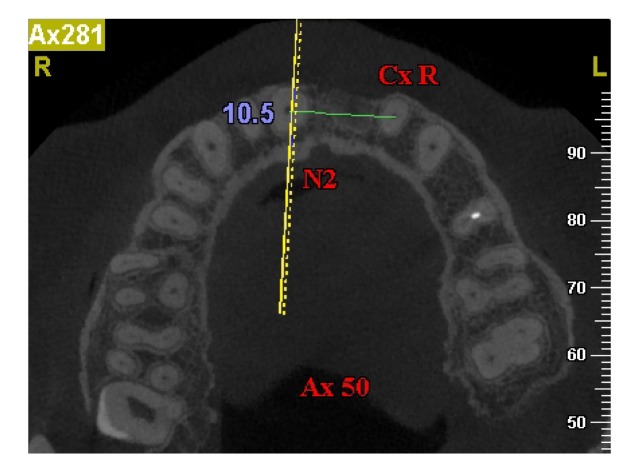
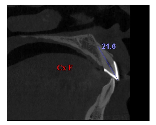
The purpose of this study was to apply cone-beam computed tomography (CBCT) to observe contour changes in human alveolar bone after tooth extraction of the maxillary central incisor and to provide original morphological evidence for aesthetic implant treatment in the maxillary anterior area. Forty patients were recruited into the study. Each patient had two CBCT scans (CBCT I and CBCT II), one taken before and one taken three months after tooth extraction of maxillary central incisor (test tooth T). A fixed anatomic reference point was used to orient the starting axial slice of the two scans. On three CBCT I axial slices, which represented the deep, middle, and shallow layers of the socket, labial and palatal alveolar bone widths of T were measured. The number of sagittal slices from the start point to the pulp centre of T was recorded. On three CBCT II axial slices, the pulp centres of extracted T were oriented according to the number of moved sagittal slices recorded in CBCT I. Labial and palatal alveolar bone widths at the oriented sites were measured. On the CBCT I axial slice which represented the middle layer of the socket, sagittal slices were reconstructed. Relevant distances of T on the sagittal slice were measured, as were the alveolar bone width and tooth length of the opposite central incisor. On the CBCT II axial slice, which represented the middle layer of the socket, relevant distances recorded in CBCT I were transferred on the sagittal slice. The height reduction of alveolar bone on labial and palatal sides was measured, as were the alveolar bone width and tooth length of the opposite central incisor at the oriented site. Intraobserver reliability assessed by intraclass correlation coefficients (ICCs) was high. Paired sample t-tests were performed. The alveolar bone width and tooth length of the opposite central incisor showed no statistical differences (P<0.05). The labial alveolar bone widths of T at the deep, middle, and shallow layers all showed statistical differences. However, no palatal alveolar bone widths showed any statistical differences. The width reduction of alveolar bone was 1.2, 1.6, and 2.7 mm at the deep, middle, and shallow layers, respectively. The height reduction of alveolar bone on labial and palatal sides of T both showed statistical differences, which was 1.9 and 1.1 mm, respectively.
Keywords: Alveolar bone contour; Cone-beam computed tomography; Maxillary central incisor.
Conflict of interest statement
All procedures followed were in accordance with the ethical standards of the responsible committee on human experimentation (institutional and national) and with the Helsinki Declaration of 1975, as revised in 2008 (5). Informed consent was obtained from all patients for being included in the study. Additional informed consent was obtained from all patients for which identifying information is included in this article.
Figures
References
MeSH terms
LinkOut - more resources
Full Text Sources
Other Literature Sources
