. 2014 Dec 4;516(7529):121-5.
doi: 10.1038/nature13980.

Piezo2 is the major transducer of mechanical forces for touch sensation in mice

Affiliations

Piezo2 is the major transducer of mechanical forces for touch sensation in mice

Sanjeev S Ranade et al. Nature. .

Abstract

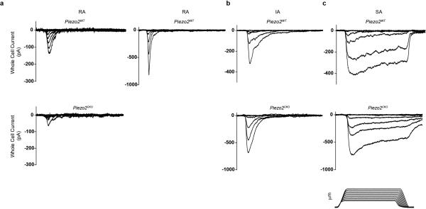
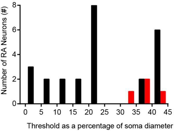
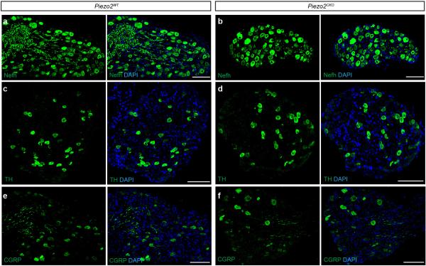
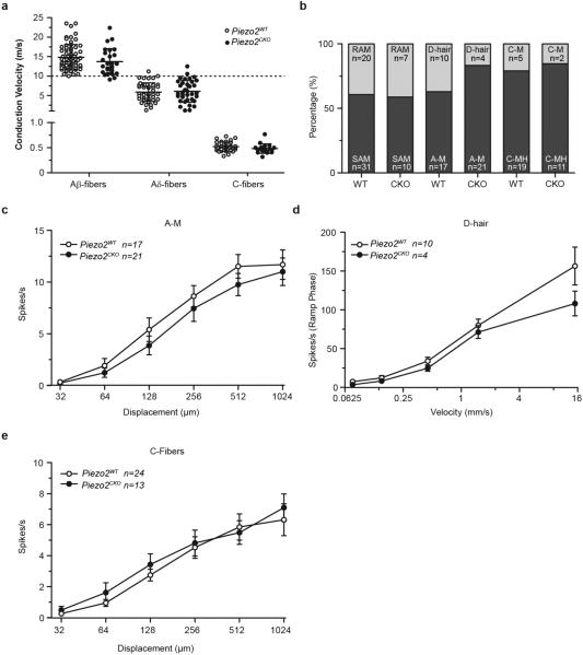
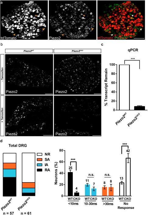
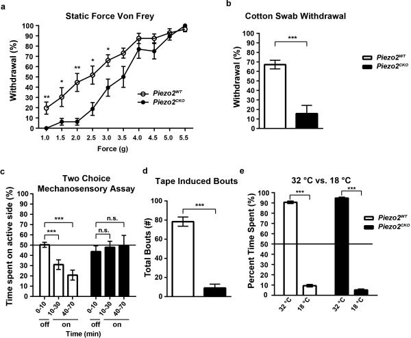
The sense of touch provides critical information about our physical environment by transforming mechanical energy into electrical signals. It is postulated that mechanically activated cation channels initiate touch sensation, but the identity of these molecules in mammals has been elusive. Piezo2 is a rapidly adapting, mechanically activated ion channel expressed in a subset of sensory neurons of the dorsal root ganglion and in cutaneous mechanoreceptors known as Merkel-cell-neurite complexes. It has been demonstrated that Merkel cells have a role in vertebrate mechanosensation using Piezo2, particularly in shaping the type of current sent by the innervating sensory neuron; however, major aspects of touch sensation remain intact without Merkel cell activity. Here we show that mice lacking Piezo2 in both adult sensory neurons and Merkel cells exhibit a profound loss of touch sensation. We precisely localize Piezo2 to the peripheral endings of a broad range of low-threshold mechanoreceptors that innervate both hairy and glabrous skin. Most rapidly adapting, mechanically activated currents in dorsal root ganglion neuronal cultures are absent in Piezo2 conditional knockout mice, and ex vivo skin nerve preparation studies show that the mechanosensitivity of low-threshold mechanoreceptors strongly depends on Piezo2. This cellular phenotype correlates with an unprecedented behavioural phenotype: an almost complete deficit in light-touch sensation in multiple behavioural assays, without affecting other somatosensory functions. Our results highlight that a single ion channel that displays rapidly adapting, mechanically activated currents in vitro is responsible for the mechanosensitivity of most low-threshold mechanoreceptor subtypes involved in innocuous touch sensation. Notably, we find that touch and pain sensation are separable, suggesting that as-yet-unknown mechanically activated ion channel(s) must account for noxious (painful) mechanosensation.


Figures

Extended Data Figure 1
Extended Data Figure 1. MA currents elicited in cultured DRG neurons from Piezo2WT and Piezo2CKO mice by poking with a blunt probe
a, Representative traces of RA currents in Piezo2WT (top) and Piezo2CKO (bottom). Piezo2WT DRG neurons show characteristic RA currents; a subpopulation can be active with apparent low thresholds (right). Piezo2CKO mice contained a few RA type cells but none were apparent low threshold mechanoreceptors. b and c, Representative traces of IA and SA currents, respectively, with no observable differences between the two genotypes. All data were low-pass filtered off line at 4KHz. Action potentials were elicited by current injection in all neurons. Piezo2WT: RA, left: 20μm diameter, 5 μm apparent threshold; RA, right: 28μm diameter, 1 μm apparent threshold; IA: 23μm diameter, 6 μm apparent threshold; SA: 20μm diameter; 2 μm apparent threshold. Piezo2CKO: RA, left: 23μm diameter, 8 μm apparent threshold; RA, right: none found; IA: 20μm diameter, 5.5 μm apparent threshold; SA: 30μm diameter, 8 μm apparent threshold. Lower right: example of probe displacement protocol (stimulus).
Extended Data Figure 2
Extended Data Figure 2. Apparent threshold analysis of Piezo2WT and Piezo2CKO DRG neurons
The smallest soma indentation eliciting a detectable MA response (apparent threshold) depends, in part, on the incremental distance applied (0.5μm) and the proportional displacement in relation to the soma diameter. The apparent thresholds of all RA responses normalized to soma diameter reveal a wide range of sensitivities of Piezo2WT DRG neurons (black) and the apparent high threshold responses of the remaining RA neurons in Piezo2CKO DRG neurons (red). The lowest apparent thresholds are observed only in Piezo2WT.
Extended Data Figure 3
Extended Data Figure 3. Expression of various markers of subpopulations of DRG neurons are similar in Piezo2WT and Piezo2CKO mice
a and b, Representative images from immunofluorescence of Nefh in DRGs from Piezo2WT (a) or Piezo2CKO (b) mice. c and d, Representative image from immunofluorescence of TH in DRGs from Piezo2WT (c) or Piezo2CKO (d) mice. e and f, Representative image from immunofluorescence of CGRP in DRGs from Piezo2WT (e) or Piezo2CKO (f) mice. All markers stained in green. Scale bars (A-F) 100μm.
Extended Data Figure 4
Extended Data Figure 4. DRG innervation of skin is unaffected in Piezo2CKO mice
a and d, Representative image of immunostaining of Krt8 (green) and Nefh (red) in Merkel cell - neurite complexes in Piezo2WT (a) and Piezo2CKO (d) glabrous skin. b, e and f, Representative image of immunostaining of S100 (green) and Nefh (red) in circumferential fibers (arrowheads) and lanceolate endings (arrows) in the hair follicle of Piezo2WT (b) and Piezo2CKO dorsal skin (e and f). c and g, Representative image of immunostaining of S100 (green) and Nefh (red) in Meissner's corpuscles in Piezo2WT (c) and Piezo2CKO (g) glabrous skin. HS, hair shaft; Bg, bulge of the hair follicle; Epi, epidermis; Der, dermis. Scale bars (a-g) 20μm.
Extended Data Figure 5
Extended Data Figure 5. Physiological properties of nociceptors are unaffected in Piezo2CKO mice
a, No change in conduction velocities of Aβ-, Aδ- and C-fiber afferents in Piezo2CKO compared Piezo2WT (Mann-Whitney Test). b, Proportions of receptor types encountered amongst Aβ-, Aδ- and C-fibers are shown. c, Stimulus response properties of A-fiber mechano-nociceptors (A-Ms) recorded in Piezo2CKO compared to Piezo2WT were not significantly different. d, D-hair receptors recorded from Piezo2CKO displayed stimulus response properties that were indistinguishable from control afferents. e, The stimulus response properties of C-fibers in Piezo2CKO were not significantly different from C-fibers recorded in control Piezo2WT mice. Data are presented as mean ± SEM, analysis for c-e with repeated measures ANOVA.
Extended Data Figure 6
Extended Data Figure 6. Development of Novel Two Choice Mechanosensory Assay
a, Schematic of instrument construction and image of instrument from top angle. b, Schematic of the instrument from the bottom angle, with the top cover removed, and photo of tactile transducers underneath the platform. c, Avoidance behavior of C57/Bl6J mice to the mechanically active side. Error bars represent SEM, n = 12 mice (c), 6 males and 6 females. ** P < 0.005, *** P <0.0001, Mann Whitney non parametric analysis.
Extended Data Figure 7
Extended Data Figure 7. Piezo2CKO mice do not show deficits in noxious mechanical or thermal stimuli or in inflammatory pain responses
a, Threshold for withdrawal response to a ramping protocol of Von Frey stimulation from low force to high in Piezo2WT (n = 9) and Piezo2CKO (n = 7) mice. b, Time to response (latency) to application of a 500g tail clip to the base of the tail in Piezo2WT (n = 6) and Piezo2CKO (n = 7) mice. c, Threshold for response to a Randall-Selitto pinching stimulus to the hind paw in Piezo2WT (n = 5) and Piezo2CKO (n = 7) mice. d, Time need to withdraw hind paw to an IR20 light (Hargreaves assay) in Piezo2WT (n = 13) and Piezo2CKO (n = 9) mice. e, ramping Von Frey protocol in baseline (before CFA injection) and 24 post CFA injection in Piezo2WT (n = 11) and Piezo2CKO (n = 9) mice. f, ramping Von Frey protocol in baseline (before BK injection) and at time points 5, 15, 15 minutes post injection in Piezo2WT (n=6) and Piezo2CKO (n=6) mice. Error bars represent SEM, all experiments performed with at least n = 2 separate cohorts of both male and female mice, ** P < 0.005, *** P <0.0001, Mann Whitney non parametric analysis.
Extended Data Figure 8
Extended Data Figure 8. Piezo1 expression and function in DRGs
a, in situ hybridization expression analysis of Piezo1 in DRG neurons relative to Piezo2. Robust expression of Piezo2 but not Piezo1 is observed, and this agrees with previously reported results using qPCR. b, siRNA for Piezo1 in cultured DRG neurons does not affect the number of mechanically sensitive neurons or ratio of RA, IA or SA type currents (number of recorded neurons in each category indicated on top of bar graphs). Data from n = 3 independent preparations, n.s. by Student's t-test). Scale bar: 100 μm (a).
Figure 1
Figure 1. Piezo2 is localized at the nerve terminals of sensory neurons that innervate the skin
a and b, representative images of immunostaining of GFP, Nefh and DAPI (blue) in the hair follicle of the Piezo2-GFP dorsal skin. GFP staining indicates localization of the Piezo2-GFP fusion protein and Nefh marks myelinated neurons. c, representative image of immunostaining of GFP, Krt8, a specific marker of Merkel cells, and DAPI (blue) in the Piezo2-GFP glabrous skin. d, representative images of immunostaining of GFP and S100, a marker of Schwann cells, in glabrous skin. Arrows mark lanceolate endings (a) and arrowheads mark circumferential fibers (b). Dashed lines demarcate epidermal-dermal junction (c and d). HS, hair shaft; Epi, epidermis; Der, dermis. Scale bars (a-d) 20μm.
Figure 2
Figure 2. AvCreERT2 mediates efficient deletion of Piezo2 and leads to specific loss of rapidly adapting (RA) mechanically activated (MA) currents in cultured DRG neurons
a, Immunostaining of DRG neurons from tamoxifen-treated AvCreERT2 x Ai9 mice for tdTomato epifluorescence and Piezo2 antibody. Arrowheads indicate the relatively few tdTomato/ Piezo2+ neurons that would presumably not be deleted in Piezo2CKO mice. b, Representative images of immunostaining using Piezo2 antibody in DRGs from AvCreERT2 x Piezo2loxP mice before and after tamoxifen treatment. c, qPCR of Piezo2 from isolated DRGs from Piezo2WT and Piezo2CKO mice with Piezo2WT values normalized at 100% (n = 3 independent experiments, P <.0001, Student's t-test). d, The proportion of DRG neurons responding with rapidly (RA, tauinact <10ms), intermediate (IA, tauinact 10-30ms), and slowly (SA, tauinact >30ms) adapting MA currents from Piezo2WT and Piezo2CKO littermates. NR, non-responsive to displacements of at least one-third cell diameter. Results from n = 3 independent experiments. Error bars represent SEM, *** P < 0.001 for RA and *** P < 0.005 for NR, Student's t-test. Scale bars (a, b) 100μm.
Figure 3
Figure 3. Piezo2 is required for mechanoreceptor function in ex vivo skin nerve preparation
a, Proportions of mechanically sensitive Aβ- and Aδ- and C-fibers in Piezo2CKO mice compared to Piezo2WT controls, Fisher's exact test ** P<0.01, one sided. b, Typical examples of SAM I mechanoreceptor responses from Piezo2CKO and Piezo2WT mice. c, Mean discharge rates (400 ms bins) during the course of ramp and hold stimuli with different onset velocities for SAM I receptors. Note the almost complete lack of dynamic ramp discharge in SAM I's recorded from Piezo2CKO mice. d, Normally, SAM I discharge rates increase with increasing ramp speed, however SAM I receptors from Piezo2CKO mice showed a strong reduction in velocity sensitivity with firing discharge increasing only marginally with increasing stimulus velocity, (repeated measures ANOVA, F=19.69, P<0.0001, with Bonferroni post-hoc test, ***P<0.001). e, Rapidly adapting afferents also displayed reduced velocity sensitivity in Piezo2CKO mutant mice (repeated measures ANOVA, F=4.36, P<0.05, with Bonferroni posthoc test, ***P<0.001). f, Mean force thresholds for nociceptor discharge. Individual nociceptors threshold were calculated by averaging the thresholds of first spikes occurring during the stimulus ramp phase for all 6 stimuli applied. (A-Ms, left; C-fibers right). Note significant elevation of mechanical thresholds for A-Ms in Piezo2CKO mice (Mann-Whitney test, * P<0.05), but no change in C-fibers mechanical thresholds. g, Discharge rates and heat thresholds to standard noxious heat ramps did not differ between polymodal C-fibers recorded from Piezo2CKO and Piezo2WT mice. Data are presented as mean ± SEM.
Figure 4
Figure 4. Piezo2CKO mice show profound and specific defects in innocuous touch sensation in multiple behavioral assays
a, Percent response to varying forces of von Frey filaments in Piezo2WT (n = 14) and Piezo2CKO mice (n = 12). b, Percent response to a sweep of cotton on the hind paw in Piezo2WT (n = 14) and Piezo2CKO mice (n = 9). Of note, 6 out of 9 Piezo2CKO mice showed zero responses to the cotton stimulus. c, Time spent on the mechanically active side over the course of 1 hour, with 10 minutes of no stimulation on either side (“off”), Piezo2WT (n = 15) and Piezo2CKO mice (n = 11). d, Number of bouts in response to a 3 cm piece of adhesive tape affixed to the back of a mouse over a 5 minute period in Piezo2WT (n = 14) and Piezo2CKO mice (n = 10). e, Percent time spent on either side of a two choice assay with one zone set at 18 °C and one zone set at 32 °C in Piezo2WT (n = 13) and Piezo2CKO mice (n = 10). Error bars represent SEM, all experiments performed with at least n = 4 separate cohorts of both male and female littermate control mice, * P < 0.05, ** P < 0.005, *** P < 0.0005, Mann Whitney non-parametric analysis.

References

    1. Abraira VE, Ginty DD. The sensory neurons of touch. Neuron. 2013;79:618–639. doi:10.1016/j.neuron.2013.07.051. - PMC - PubMed
    1. Arnadottir J, Chalfie M. Eukaryotic mechanosensitive channels. Annu Rev Biophys. 2010;39:111–137. doi:10.1146/annurev.biophys.37.032807.125836. - PubMed
    1. Coste B, et al. Piezo1 and Piezo2 are essential components of distinct mechanically activated cation channels. Science. 2010;330:55–60. doi:10.1126/science.1193270. - PMC - PubMed
    1. Woo SH, et al. Piezo2 is required for Merkel-cell mechanotransduction. Nature. 2014 doi:10.1038/nature13251. - PMC - PubMed
    1. Maksimovic S, et al. Epidermal Merkel cells are mechanosensory cells that tune mammalian touch receptors. Nature. 2014 doi:10.1038/nature13250. - PMC - PubMed

Publication types