. 2015 Feb;89(4):2112-20.
doi: 10.1128/JVI.02245-14. Epub 2014 Dec 3.

Regulatory T cells resist virus infection-induced apoptosis

Affiliations

Regulatory T cells resist virus infection-induced apoptosis

Jenny W Che et al. J Virol. 2015 Feb.

Abstract

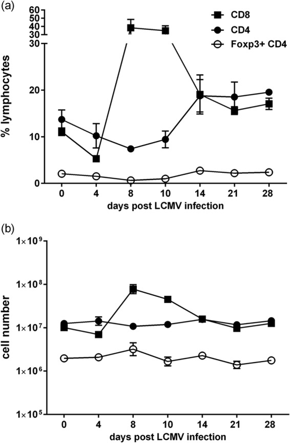
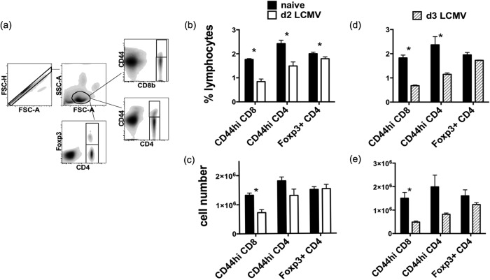
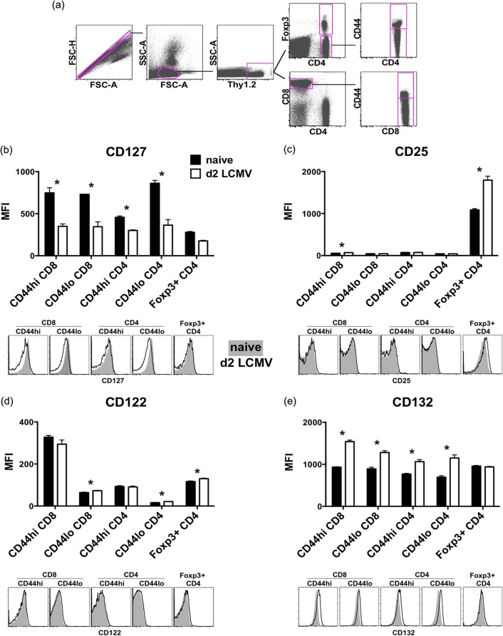
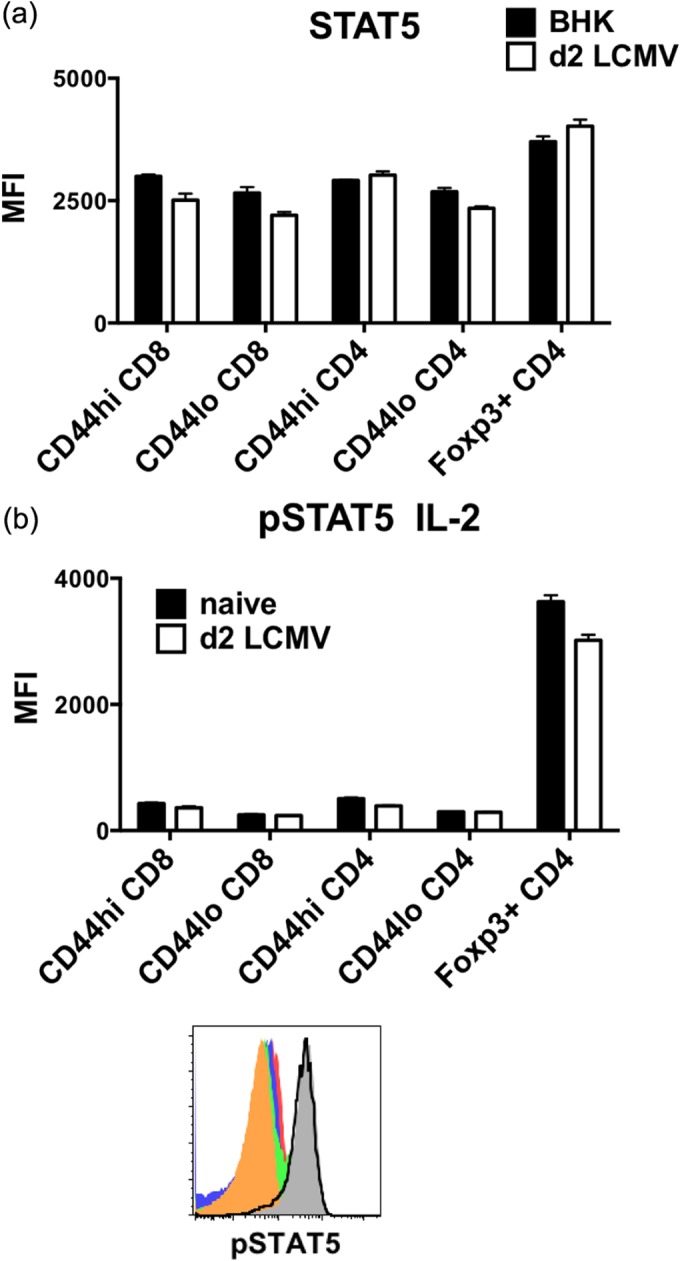
Regulatory T (Treg) cells are important in the maintenance of self-tolerance, and the depletion of Treg cells correlates with autoimmune development. It has been shown that type I interferon (IFN) responses induced early in the infection of mice can drive memory (CD44hi) CD8 and CD4 T cells into apoptosis, and we questioned here whether the apoptosis of CD44-expressing Treg cells might be involved in the infection-associated autoimmune development. Instead, we found that Treg cells were much more resistant to apoptosis than CD44hi CD8 and CD4 T cells at days 2 to 3 after lymphocytic choriomeningitis virus infection, when type I IFN levels are high. The infection caused a downregulation of the interleukin-7 (IL-7) receptor, needed for survival of conventional T cells, while increasing on Treg cells the expression of the high-affinity IL-2 receptor, needed for STAT5-dependent survival of Treg cells. The stably maintained Treg cells early during infection may explain the relatively low incidence of autoimmune manifestations among infected patients.

Importance: Autoimmune diseases are controlled in part by regulatory T cells (Treg) and are thought to sometimes be initiated by viral infections. We tested the hypothesis that Treg may die off at early stages of infection, when virus-induced factors kill other lymphocyte types. Instead, we found that Treg resisted this cell death, perhaps reducing the tendency of viral infections to cause immune dysfunction and induce autoimmunity.


Figures

FIG 1
FIG 1
Relatively stable Foxp3+ CD4+ Treg cell numbers during LCMV infection. C57BL/6 mice were infected i.p. with 5 × 10⁴ PFU of LCMV strain Armstrong, and the spleen was examined every 4 days over 4 weeks for the frequency (a) and total number (b) of CD8 T, Foxp3+ CD4+ Treg, and Foxp3-negative CD4 T cells. Each time point represents 3 to 14 mice.
FIG 2
FIG 2
Foxp3+ CD4+ Treg cells resist loss after LCMV infection in comparison to CD44hi CD8 and CD44hi Foxp3-negative CD4 T cells. Foxp3-GFP mice were infected i.p. with 5 × 10⁴ PFU LCMV. Two (b and c) or 3 (d and e) days later, surface-stained splenocytes from infected and uninfected (naive) mice were analyzed by flow cytometry, with gating as shown in panel a. (b and d) The frequencies of CD8 T cells and Foxp3+ CD4+ Treg cells were obtained by direct gating, while the frequency of Foxp3-negative CD4 T cells was calculated by Boolean gating in FlowJo. (c and e) The absolute numbers of the different populations were then calculated from the total live splenocyte counts. (b and c) Combined data from 3 separate experiments (n = 9). (d and e) Representative data from seven separate experiments (n = 3). *, statistical significance (P < 0.05) between naive and LCMV-infected groups.
FIG 3
FIG 3
Foxp3+ CD4+ Treg cells are less TUNEL positive than CD44hi Foxp3-negative CD4 and CD44hi CD8 T cells in the spleen at day 2 following LCMV infection. Foxp3-GFP mice were infected i.p. with 5 × 10⁴ PFU LCMV. Splenocytes harvested at 2 days postinfection were stained with TUNEL after incubating in medium at 37°C for 5 h. (a) Histograms of TUNEL staining intensity of CD4, CD8, and Treg cells from a naive animal and a day 2 LCMV-infected animal in a representative experiment. (b) Combined data from 3 separate experiments (total n = 9). *, statistical significance (P < 0.05) between naïve mice and mice at day 2 following LCMV infection. (c) A representative panel of dot plots showing TUNEL reactivity versus CD44 staining among CD4 T cells, CD8 T cells, and Treg cells.
FIG 4
FIG 4
Foxp3+ CD4+ Treg cells are less annexin V positive than CD8 and Foxp3-negative CD4 T cells in the spleen at day 2 following LCMV infection. Foxp3-GFP mice were infected i.p. with 5 × 10⁴ PFU LCMV. Splenocytes harvested at 2 days postinfection were stained ex vivo with Live/Dead Fixable stain and annexin V. (a) Histograms of annexin V staining intensity of live CD4, CD8, and Treg cells from a naive animal and a day 2 LCMV-infected animal in a representative experiment. (b) Data are from a representative of three separate experiments (n = 3). *, statistical significance (P < 0.05) between naive cells and cells at day 2 following LCMV infection. (c) Representative panel of dot plots showing annexin V reactivity versus CD44 staining among CD4 and CD8 T cells and Treg cells.
FIG 5
FIG 5
Foxp3+ CD4+ Treg cells are less apoptotic than CD44hi Foxp3-negative CD4 and CD8 T cells in spleen at day 3 following LCMV infection. Foxp3-GFP mice were infected i.p. with 5 × 10⁴ PFU LCMV. Splenocytes harvested at 3 days postinfection were stained with TUNEL after incubating in medium at 37°C for 5 h (a) or ex vivo with Live/Dead Fixable stain and annexin V (b). Data are representative of at least three separate experiments (n = 3). *, statistical significance (P < 0.05) between naive cells and cells at day 3 following LCMV infection.
FIG 6
FIG 6
The IL-7 receptor is downregulated on T cells, while the high-affinity IL-2 receptor remains on Foxp3+ CD4+ Treg cells after LCMV infection. Foxp3-GFP mice were infected i.p. with 5 × 10⁴ PFU LCMV. Splenocytes harvested at 2 days postinfection were stained ex vivo for CD127 (b), CD25 (c), CD122 (d), and CD132 (e), and the T cell populations were analyzed by flow cytometry (a). Data are representative of at least three separate experiments showing geometric mean fluorescence intensity between naive mice and mice at day 2 following LCMV infection (n = 3). *, statistical significance (P < 0.05) between naive mice and mice at day 2 following LCMV infection (n = 3). Histograms of a representative animal from each group were overlaid for visual comparison.
FIG 7
FIG 7
Foxp3+ CD4+ Treg cells display the highest level of STAT5 expression and respond well to IL-2 stimulation in vitro even after LCMV infection. Foxp3-GFP mice were infected i.p. with 5 × 10⁴ PFU LCMV. (a) Splenocytes harvested at 2 days postinfection were stained ex vivo for STAT5. (b) Phosphorylation of STAT5 (pSTAT5) was assessed among CD44hi CD8 (solid red), CD44lo CD8 (solid blue), CD44hi Foxp3-negative CD4 (solid green), CD44lo Foxp3-negative CD4 (solid orange), and Foxp3+ CD4 (solid gray) T cells from a representative naive animal and Foxp3+ CD4 T cells (black line) from a day 2 LCMV-infected animal after stimulating splenocytes with 5 ng/ml IL-2 for 15 min at 37°C. Data are representative of two separate experiments showing geometric mean fluorescence intensity between BHK sham control or naive versus day 2 LCMV-infected groups of 2 or 3 mice.

References

    1. Zheng Y, Josefowicz SZ, Kas A, Chu T-T, Gavin MA, Rudensky AY. 2007. Genome-wide analysis of Foxp3 target genes in developing and mature regulatory T cells. Nature 445:936–940. doi: 10.1038/nature05563. - DOI - PubMed
    1. Brunkow ME, Jeffery EW, Hjerrild KA, Paeper B, Clark LB, Yasayko S-A, Wilkinson JE, Galas D, Ramsdell SFZF. 2001. Disruption of a new forkhead/winged-helix protein, scurfin, results in the fatal lymphoproliferative disorder of the scurfy mouse. Nat Genet 27:68–73. doi: 10.1038/83784. - DOI - PubMed
    1. Godfrey VL, Wilkinson JE, Russell LB. 1991. X-linked lymphoreticular disease in the scurfy (sf) mutant mouse. Am J Pathol 138:1379–1387. - PMC - PubMed
    1. Feuerer M, Shen Y, Littman DR, Benoist C, Mathis D. 2009. How punctual ablation of regulatory T cells unleashes an autoimmune lesion within the pancreatic islets. Immunity 31:654–664. doi: 10.1016/j.immuni.2009.08.023. - DOI - PMC - PubMed
    1. Lahl K, Loddenkemper C, Drouin C, Freyer J, Arnason J, Eberl G, Hamann A, Wagner H, Huehn J, Sparwasser T. 2007. Selective depletion of Foxp3+ regulatory T cells induces a scurfy-like disease. J Exp Med 204:57–63. doi: 10.1084/jem.20061852. - DOI - PMC - PubMed

Publication types

MeSH terms